Osborne Head & Neck Institute | Los Angeles
Salivary Gland Diagnostics
Imaging, Biopsy & Advanced Evaluation
Ryan F. Osborne, MD | Fellowship-Trained Head & Neck Surgical Oncologist
Medically Reviewed By: OHNI Clinical Staff · Last Updated: November 3, 2026
Foundation
Principles of Salivary Gland Diagnostics
Precision Before Treatment
Definitive management decisions should only be made after thorough diagnostic evaluation. Misclassification can lead to overtreatment or delayed care.
Multimodal Assessment
No single diagnostic tool is sufficient in isolation. Optimal evaluation combines clinical examination, imaging, tissue sampling, and functional assessment.
Minimally Invasive First
Whenever possible, diagnostic strategies prioritize low-risk, minimally invasive techniques that provide high-yield information.
First Step
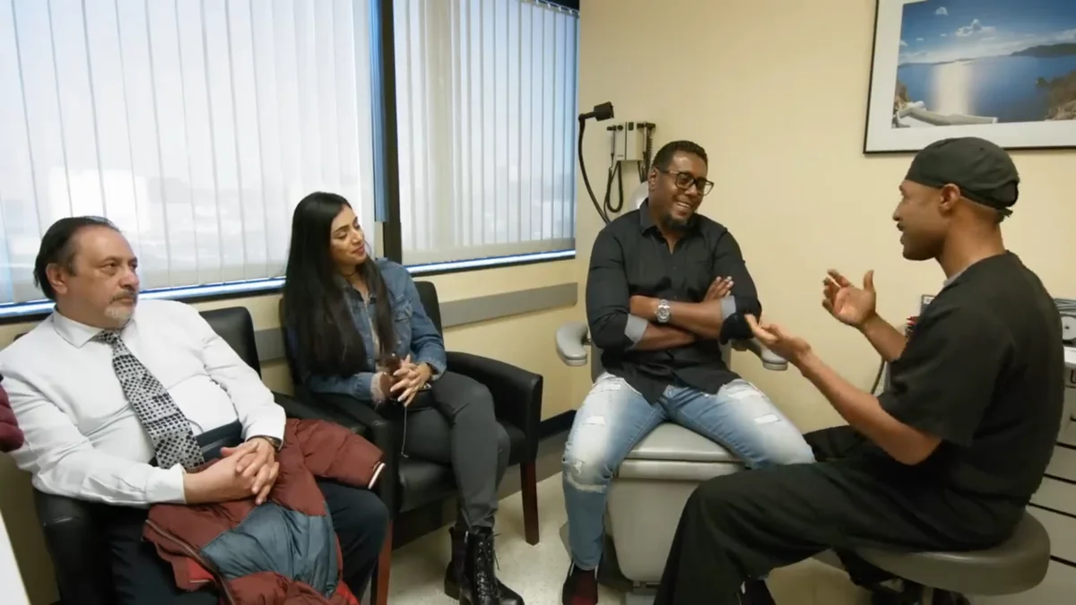
Clinical Evaluation
The diagnostic process begins with a detailed clinical assessment. Clinical findings guide selection of appropriate imaging and further testing.
History
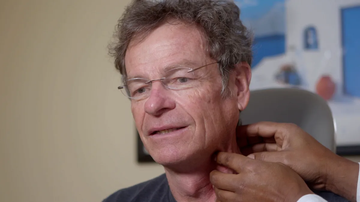
Physical Examination
Advanced Imaging
Imaging of Salivary Gland Disorders
Imaging plays a central role in defining disease extent, guiding biopsy, and planning treatment.
Ultrasound
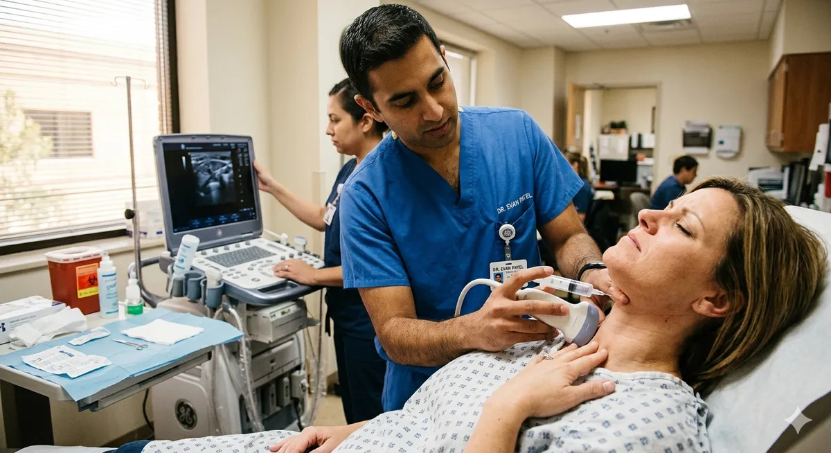
First-line imaging for superficial lesions. Real-time, non-invasive, and cost-effective. Useful for guiding fine needle aspiration.
MRI
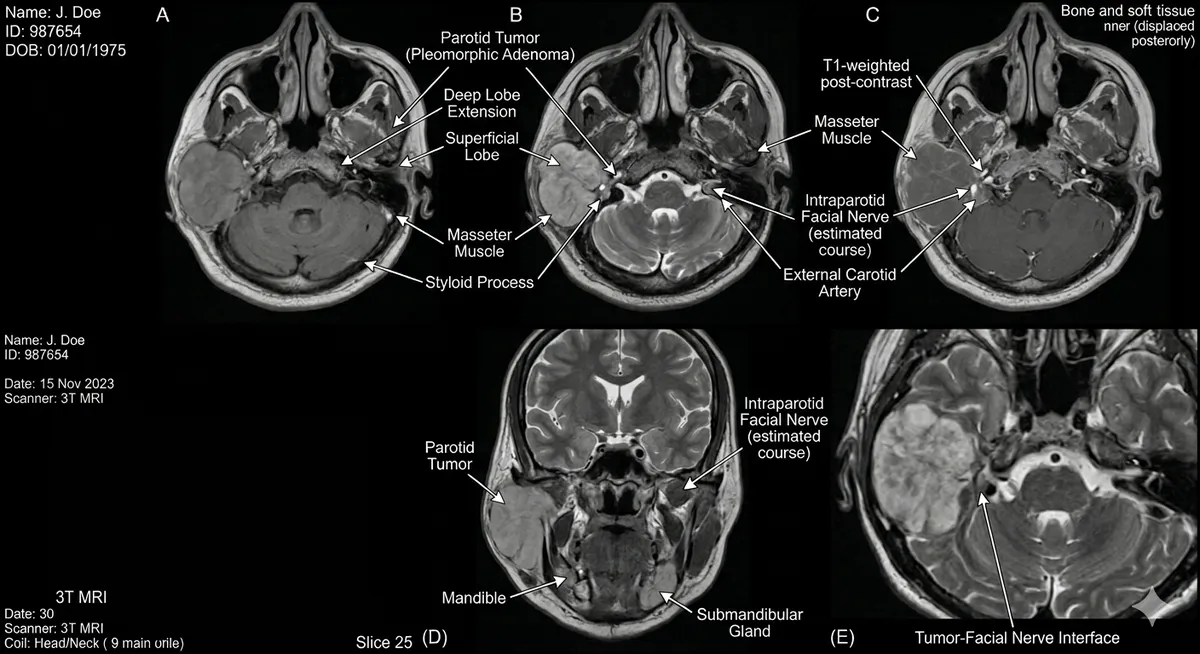
Superior soft tissue resolution. Essential for evaluating parotid tumors and deep lobe disease. Provides detailed relationship to the facial nerve.
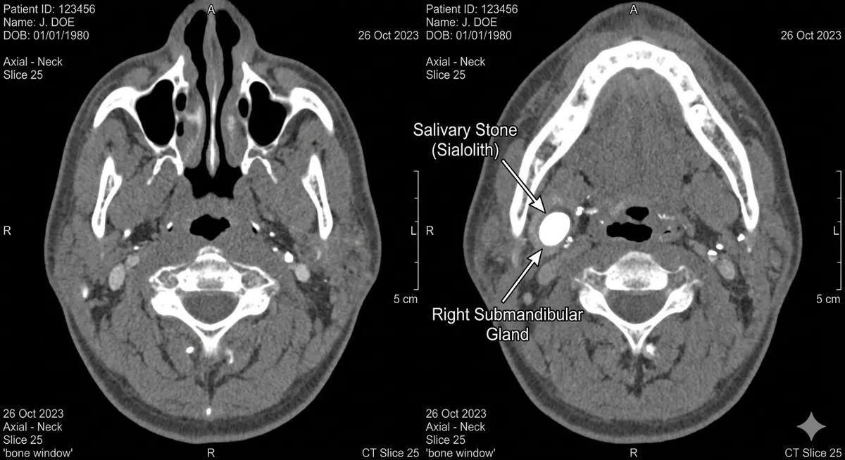
CT Scan
Useful for detecting calcifications and salivary stones. Helpful in evaluating bony involvement and acute infection.
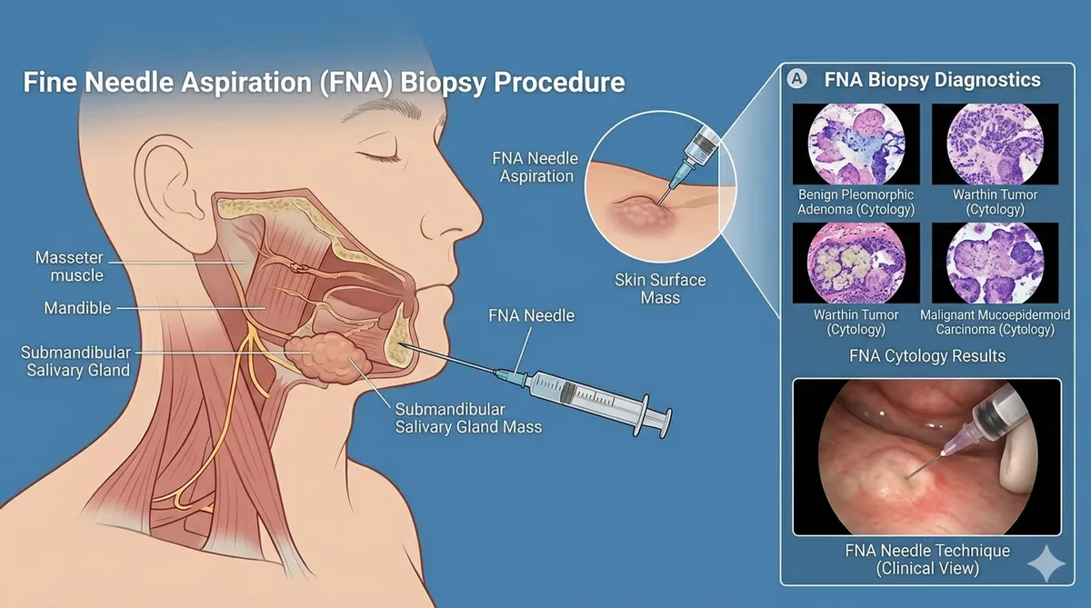
Fine Needle Aspiration (FNA)
FNA biopsy is a key diagnostic tool for evaluating salivary gland masses. Minimally invasive, it provides cytologic information and helps distinguish benign from malignant disease.
- Minimally invasive
- Provides cytologic information
- Helps distinguish benign from malignant disease
Sialendoscopy
Direct visualization of the salivary ductal system. Identifies stones, strictures, and inflammatory changes. Can be combined with therapeutic intervention.
- Identifies stones, strictures, and inflammatory changes
- Can be combined with therapeutic intervention
- Minimally invasive approach
Condition-Specific
Diagnostic Pathways by Condition
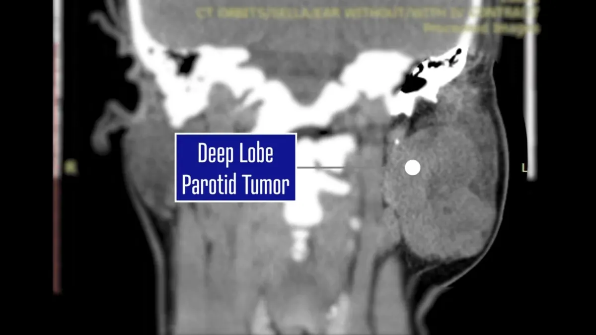
Evaluation of Parotid Masses
Clinical examination
MRI for tumor characterization
FNA biopsy for cytologic assessment
Evaluation of Obstructive Symptoms
Ultrasound or CT for stone detection
Sialendoscopy for confirmation and treatment
Evaluation of Inflammatory Disease
Clinical assessment
Imaging to identify obstruction or chronic changes
Consider autoimmune workup when indicated
Evaluation of Dry Mouth
Functional testing of salivary output
Assessment for systemic or medication-related causes
Our Approach
Diagnostic Philosophy
Avoid Unnecessary Intervention
Accurate diagnosis reduces the risk of unnecessary surgery or inappropriate treatment.
Early and Accurate Identification
Timely diagnosis improves outcomes, particularly in neoplastic and obstructive disease.
Integration with Treatment Pathways
Diagnostics are not isolated steps but part of a continuous care pathway leading to definitive management.
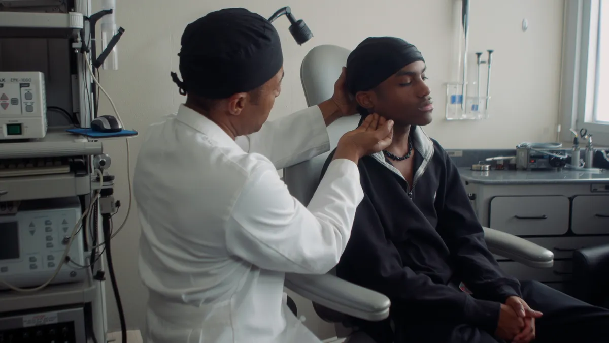
Don't Wait
When to Seek Specialist Evaluation
Early evaluation allows for precise diagnosis and expands available treatment options. Seek evaluation if you have:
A persistent mass in the salivary gland region
Recurrent swelling or pain
Unexplained dry mouth
Facial weakness or asymmetry
PATIENT EDUCATION
Salivary Gland Diagnostics FAQs
MRI is generally the preferred imaging modality for salivary gland evaluation. It provides excellent soft tissue detail and helps distinguish between benign and malignant tumors. CT and ultrasound also play important roles depending on the clinical scenario.
FNA is a minimally invasive procedure where a thin needle is used to extract cells from a salivary gland mass for pathological analysis. It helps determine whether a mass is benign or malignant before surgery.
In most cases, FNA biopsy is recommended to guide treatment planning. However, the decision depends on imaging characteristics, clinical findings, and the overall clinical context.
Modern imaging combined with FNA biopsy provides high diagnostic accuracy. MRI can identify tumor location, size, and characteristics, while FNA helps confirm the pathological diagnosis.
Sialography is an imaging technique where contrast dye is injected into the salivary duct to visualize the ductal system. It is useful for evaluating duct obstruction, strictures, and chronic inflammatory conditions.
Why Choose OHNI
Not Big Medicine | Personalized, Global Expertise
Many patients assume the best care is found at large, well-known institutions. While these centers offer excellent care, they often operate within standardized systems shaped by regional training models.
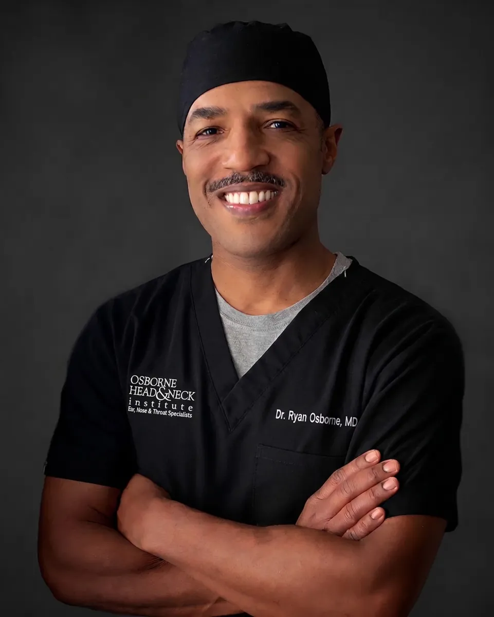
At OHNI, Dr. Ryan Osborne offers something fundamentally different: a truly global perspective combined with highly personalized surgical care with experience across five continents, tailoring each procedure to the individual patient, not the system.
Global Perspective
Experience across five continents, applying diverse surgical philosophies and techniques beyond standard U.S. training.
Precision & Preservation
Specialized techniques like Extra-Capsular Dissection designed to minimize invasiveness while protecting the facial nerve.
Patient-First Care
Direct surgeon access, individualized treatment planning, and a focus on both medical outcomes and quality of life.
Proven Outcomes
Patients consistently highlight clear communication, confident surgical decisions, and excellent functional results.
Precision Diagnostics for Salivary Gland Disorders
Through structured salivary gland diagnostics, it is possible to accurately classify disease, guide treatment decisions, and optimize patient outcomes.
Once a diagnosis is confirmed and surgery is recommended, Parotidectomy Recovery and Parotid Surgery Risks explain the next steps.
Schedule a Diagnostic Consultation
Contact the Osborne Head & Neck Institute for expert diagnostic evaluation of salivary gland disorders.