Osborne Head & Neck Institute | Los Angeles
Fine Needle Aspiration (FNA) Biopsy
Minimally Invasive Diagnostic Tool for Salivary Gland Masses
Ryan F. Osborne, MD | Fellowship-Trained Head & Neck Surgical Oncologist
Medically Reviewed By: OHNI Clinical Staff · Last Updated: November 3, 2026
Overview
What Is Fine Needle Aspiration (FNA)?
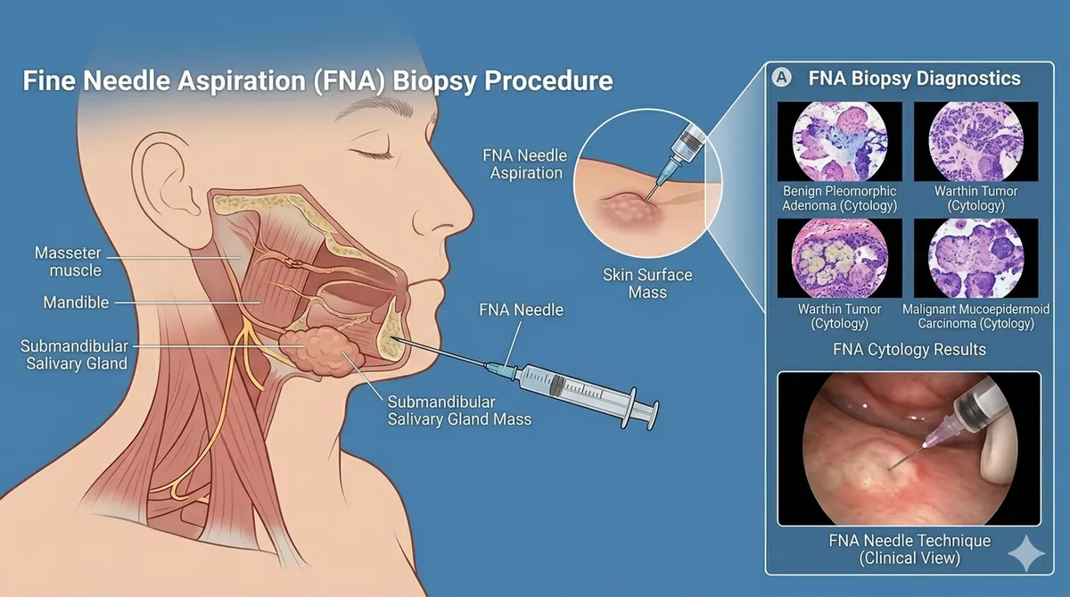
Fine needle aspiration is a diagnostic procedure in which a thin, hollow needle, typically 23–25 gauge, is inserted into a mass to extract cellular material for microscopic examination.2 The collected cells are then stained and analyzed by a cytopathologist, allowing clinicians to assess the nature of the lesion without the need for a more invasive surgical biopsy.
This technique is often referred to as fine-needle aspiration cytology (FNAC), emphasizing that the analysis focuses on individual cells rather than tissue architecture.2 FNA can be performed in an outpatient setting, often without anesthesia for superficial lesions, and is associated with minimal discomfort and a rapid recovery time.3
Clinical Role
Role of FNA in Evaluating Salivary Gland Masses
Differentiate Benign vs. Malignant
Distinguish between benign and malignant tumors before any surgical intervention.
Identify Inflammation
Detect inflammatory or infectious processes that may mimic neoplastic disease.
Detect Metastatic Disease
Identify metastatic disease or lymphoma affecting the salivary glands.
Guide Treatment Planning
Inform treatment planning and surgical decision-making with cytologic data.
Clinical and radiologic evaluation alone is often insufficient to distinguish benign from malignant lesions.1 Therefore, FNA provides a critical layer of diagnostic clarity before proceeding to surgery or additional interventions.
Cytology
What FNA Reveals
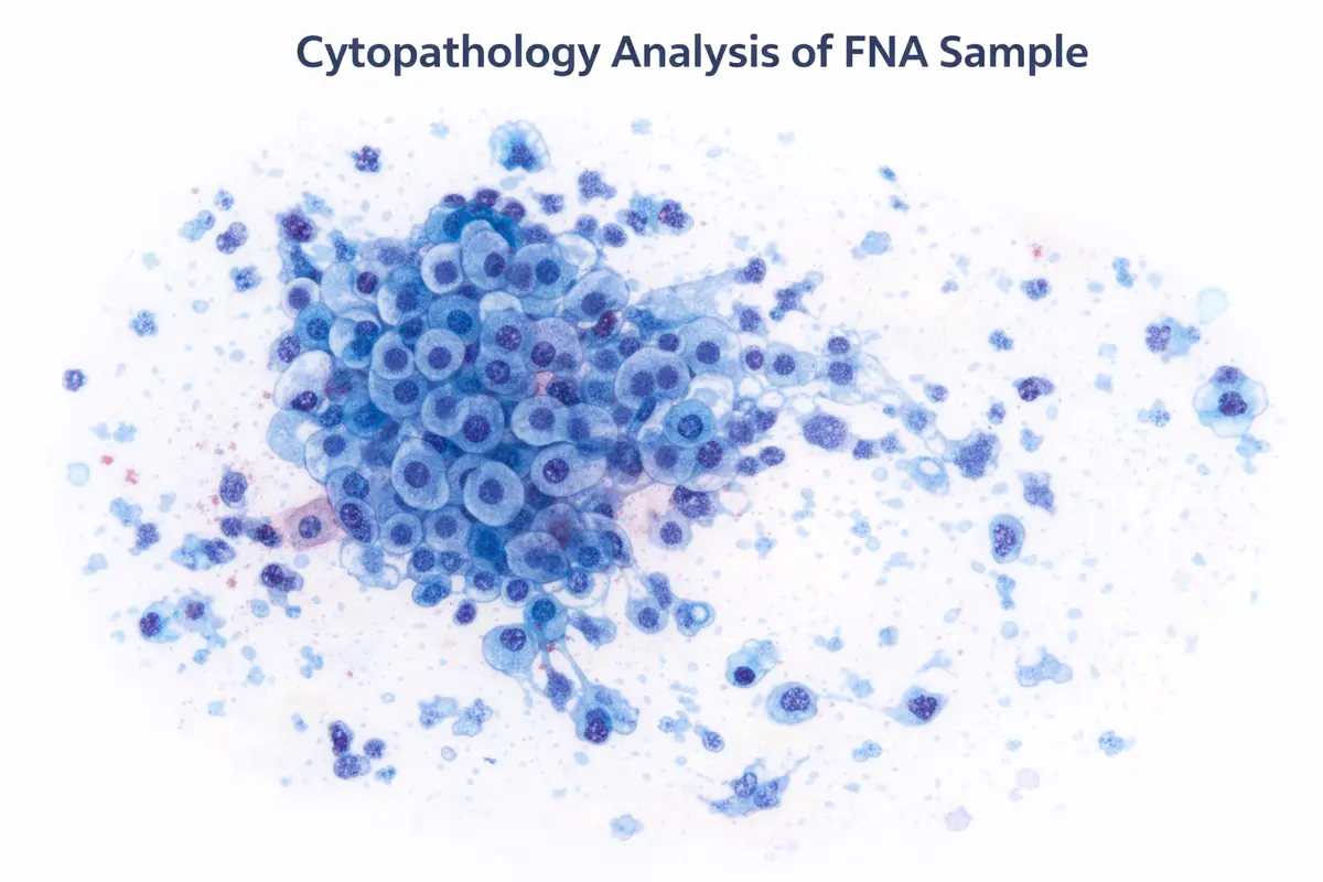
FNA biopsy provides cytologic information, meaning it allows examination of individual cells rather than whole tissue structures. This information can reveal:
- Cellular morphology (shape, size, nuclear features)
- Presence of atypia or malignancy
- Inflammatory or reactive changes
- Tumor type (in some cases)
The primary goal of cytologic evaluation is to determine whether a lesion is non-neoplastic (e.g., infection or inflammation), a benign neoplasm (e.g., pleomorphic adenoma), or a malignant tumor (e.g., mucoepidermoid carcinoma). This distinction is critical because it directly influences management strategies.5
Lesion Classification
Non-Neoplastic
Infection, inflammation, reactive changes
Benign Neoplasm
Pleomorphic adenoma, Warthin tumor
Malignant Tumor
Mucoepidermoid carcinoma, adenoid cystic carcinoma
Evidence
Diagnostic Accuracy of FNA Biopsy
FNA biopsy is widely regarded as a reliable diagnostic tool with high accuracy, though results can vary depending on technique and expertise.6
67–93%
Sensitivity
98–100%
Specificity
95–96%
Overall Accuracy
High specificity means that when FNA indicates malignancy, it is highly likely to be correct. However, sensitivity can vary, meaning that some malignant lesions may not be detected (false negatives), particularly if sampling is inadequate or the tumor is heterogeneous.7
Benefits
Advantages of FNA Biopsy
Rapid & Cost-Effective
FNA is relatively inexpensive and can provide results quickly, allowing for timely clinical decisions.
Outpatient Procedure
Most FNA biopsies are performed in an office or clinic without hospitalization.
Reduced Need for Surgery
In some cases, FNA can help avoid unnecessary surgery for benign or non-neoplastic conditions.
Preoperative Planning
Knowing whether a tumor is benign or malignant helps surgeons plan the extent of surgery and counsel patients.
Safe & Well-Tolerated
The procedure has a low complication rate and minimal recovery time.
Considerations
Limitations & Challenges of FNA
Sampling Error
If insufficient or non-representative cells are collected, the result may be inconclusive or inaccurate.
Cytologic Overlap
Many salivary gland tumors have overlapping cellular features, making precise diagnosis challenging.
Limited Tumor Grading
FNA may not always provide enough information to determine tumor grade or exact subtype.
Operator & Interpreter Dependence
Accuracy depends heavily on the skill of the clinician performing the biopsy and the pathologist interpreting the results.
False Negatives
A negative result does not always rule out malignancy, particularly in cases with inadequate sampling.
Indications
When Is FNA Recommended?
FNA biopsy is typically recommended in the following situations:8
- A new or unexplained salivary gland mass
- A mass with suspicious clinical or imaging features
- Evaluation of recurrent or persistent swelling
- Suspected metastatic disease or lymphoma
- Patients who are poor surgical candidates
It is particularly useful when the diagnosis will influence treatment decisions or surgical planning.
FNA vs. Core Needle Biopsy
Fine Needle Aspiration
- Less invasive
- Faster and easier to perform
- Provides cytologic (cell-level) information
Core Needle Biopsy
- More invasive
- Provides histologic (tissue-level) architecture
- Often higher diagnostic accuracy for tumor subtyping
Some studies suggest CNB may offer superior diagnostic detail, but FNA remains a valuable first-line tool due to its safety and convenience.6
The Procedure
What Patients Can Expect
The FNA procedure is straightforward and typically takes only a few minutes.2
Preparation
The skin over the biopsy site is cleaned, and local anesthesia may be applied if needed.
Needle Insertion
A fine needle is inserted into the mass, often guided by palpation or ultrasound.
Cell Aspiration
Cells are withdrawn using suction and placed onto slides for analysis.
Multiple Passes
Several samples may be taken to ensure adequate material for accurate diagnosis.
Post-Procedure
Patients can usually resume normal activities immediately, with minimal discomfort.
Advanced Technique
Ultrasound-Guided FNA
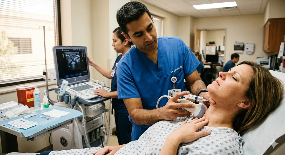
Ultrasound guidance has improved the accuracy of FNA by allowing precise needle placement, especially in small or deep lesions. Studies show that ultrasound-guided FNA can achieve diagnostic accuracy rates approaching 95% when adequate samples are obtained.7
- Improved sampling accuracy
- Reduced risk of complications
- Better visualization of lesion characteristics
Clinical Impact
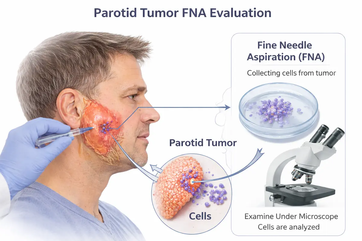
FNA in Parotid Tumors
In parotid gland tumors, the most common salivary gland neoplasms, FNA plays a particularly important role. Most parotid masses are benign, but a significant minority are malignant.9 FNA helps:
- Avoid overtreatment of benign lesions
- Identify malignancy early
- Guide decisions about facial nerve preservation
- Inform patients about prognosis and treatment options
Even when surgery is ultimately required, FNA provides valuable preoperative information that improves patient counseling and surgical planning.
If surgery is recommended, Parotidectomy Recovery explains what to expect afterward, and Parotid Surgery Risks covers possible complications.
Emerging Advances
The Future of FNA in Salivary Gland Diagnosis
Advances in cytopathology and molecular diagnostics are enhancing the value of FNA biopsy. These innovations aim to increase diagnostic accuracy and provide more precise tumor classification.10
References
- Schmidt RL, Hall BJ, Wilson AR, Layfield LJ. A systematic review and meta-analysis of the diagnostic accuracy of fine-needle aspiration cytology for salivary gland lesions. American Journal of Clinical Pathology. 2011.
- Orell SR, Sterrett GF, Whitaker D. Fine Needle Aspiration Cytology. 5th ed. Elsevier; 2012.
- Kocjan G. Diagnostic Cytopathology Essentials. Churchill Livingstone; 2010.
- Seethala RR, Stenman G. Update from the 4th edition of the WHO classification of head and neck tumours: tumors of the salivary gland. Head and Neck Pathology. 2017.
- Rossi ED, Wong LQ, Bizzarro T, et al. The Milan System for Reporting Salivary Gland Cytopathology. Acta Cytologica. 2018.
- Novoa E, Gürtler N, Arnoux A, Kraft M. Role of fine-needle aspiration cytology and core needle biopsy in the assessment of salivary gland lesions. Head & Neck. 2016.
- Howlett DC, Harper B, Quante M, et al. Diagnostic adequacy and accuracy of ultrasound-guided fine needle aspiration cytology in salivary gland lesions. American Journal of Roentgenology. 2007.
- Pynnonen MA, Gillespie MB, Roman B, et al. Clinical practice guideline: evaluation of the neck mass in adults. Otolaryngology–Head and Neck Surgery. 2017.
- Speight PM, Barrett AW. Salivary gland tumours. Oral Diseases. 2002.
- Griffith CC, Pai RK, Schneider F, et al. Salivary gland tumor cytology: Milan system and emerging molecular tools. Biomedicines. 2023.
Patient Education
Frequently Asked Questions
Most patients experience minimal discomfort during an FNA biopsy. The needle used is very thin, and the procedure is quick. Some patients report a brief pinching sensation, and mild soreness afterward is possible but usually resolves within a day or two.
Results are typically available within a few days, depending on the laboratory and whether additional testing is required. In some cases, preliminary results may be available sooner if a cytopathologist evaluates the sample on-site.
FNA biopsy is highly accurate, particularly for distinguishing benign from malignant lesions. However, it may not always provide a definitive diagnosis or tumor subtype, and additional testing or surgery may be required in certain cases.
If results are inconclusive or insufficient, the procedure may be repeated, often with ultrasound guidance. Alternatively, a core needle biopsy or surgical biopsy may be recommended for further evaluation.
Yes, FNA is considered very safe for evaluating parotid gland masses. Complications are rare and typically minor, such as slight bruising or swelling. The risk of tumor spread from FNA is extremely low.
Why Choose OHNI
Not Big Medicine | Personalized, Global Expertise
Many patients assume the best care is found at large, well-known institutions. While these centers offer excellent care, they often operate within standardized systems shaped by regional training models.
At OHNI, Dr. Ryan Osborne offers something fundamentally different: a truly global perspective combined with highly personalized surgical care with experience across five continents, tailoring each procedure to the individual patient, not the system.
Global Perspective
Experience across five continents, applying diverse surgical philosophies and techniques beyond standard U.S. training.
Precision & Preservation
Specialized techniques like Extra-Capsular Dissection designed to minimize invasiveness while protecting the facial nerve.
Patient-First Care
Direct surgeon access, individualized treatment planning, and a focus on both medical outcomes and quality of life.
Proven Outcomes
Patients consistently highlight clear communication, confident surgical decisions, and excellent functional results.
Need a Salivary Gland Biopsy Evaluation?
Our specialists provide expert FNA biopsy and comprehensive salivary gland diagnostics. Schedule a consultation to get accurate answers about your condition.
Become a Patient
Contact the Osborne Head & Neck Institute for expert evaluation and surgical planning.