Osborne Head & Neck Institute | Los Angeles
Salivary Gland Stones
Sialolithiasis, Diagnosis & Minimally Invasive Treatment
Ryan F. Osborne, MD | Fellowship-Trained Head & Neck Surgical Oncologist
Medically Reviewed By: OHNI Clinical Staff · Last Updated: November 3, 2026
Understanding the Condition
Overview of Salivary Stone Disease
Salivary gland stones (sialolithiasis) are hardened mineral deposits that form inside a salivary gland or its duct and can block the flow of saliva. Salivary stones most frequently affect the submandibular glands, although the parotid glands may also be involved. Stone formation is influenced by factors such as saliva composition, duct anatomy, and flow dynamics.
Obstruction leads to increased pressure within the gland, resulting in inflammation and progressive dysfunction if untreated.
Classification
Types of Salivary Gland Stones
Submandibular Salivary Gland Stones
The majority of salivary stones occur in the submandibular gland due to its more viscous saliva and upward ductal flow.
Parotid Salivary Gland Stones
Parotid stones are less common but often more technically challenging due to smaller duct size and branching anatomy.
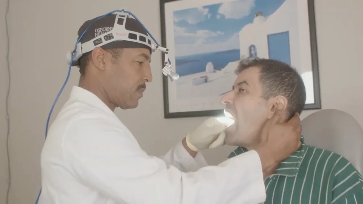
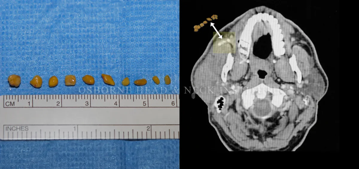
Dr. Osborne examining a patient with a salivary gland stone
Signs & Symptoms
Symptoms of Salivary Stones
Patients commonly present with classic obstructive symptoms that follow a cyclical pattern related to salivary stimulation:
Swelling that worsens during eating
Intermittent or persistent gland pain
Decreased salivary flow
Recurrent infections or tenderness
Precision Diagnostics
Diagnostic Evaluation
Accurate diagnosis requires integration of clinical findings and imaging.
Imaging Modalities
- Ultrasound for initial detection
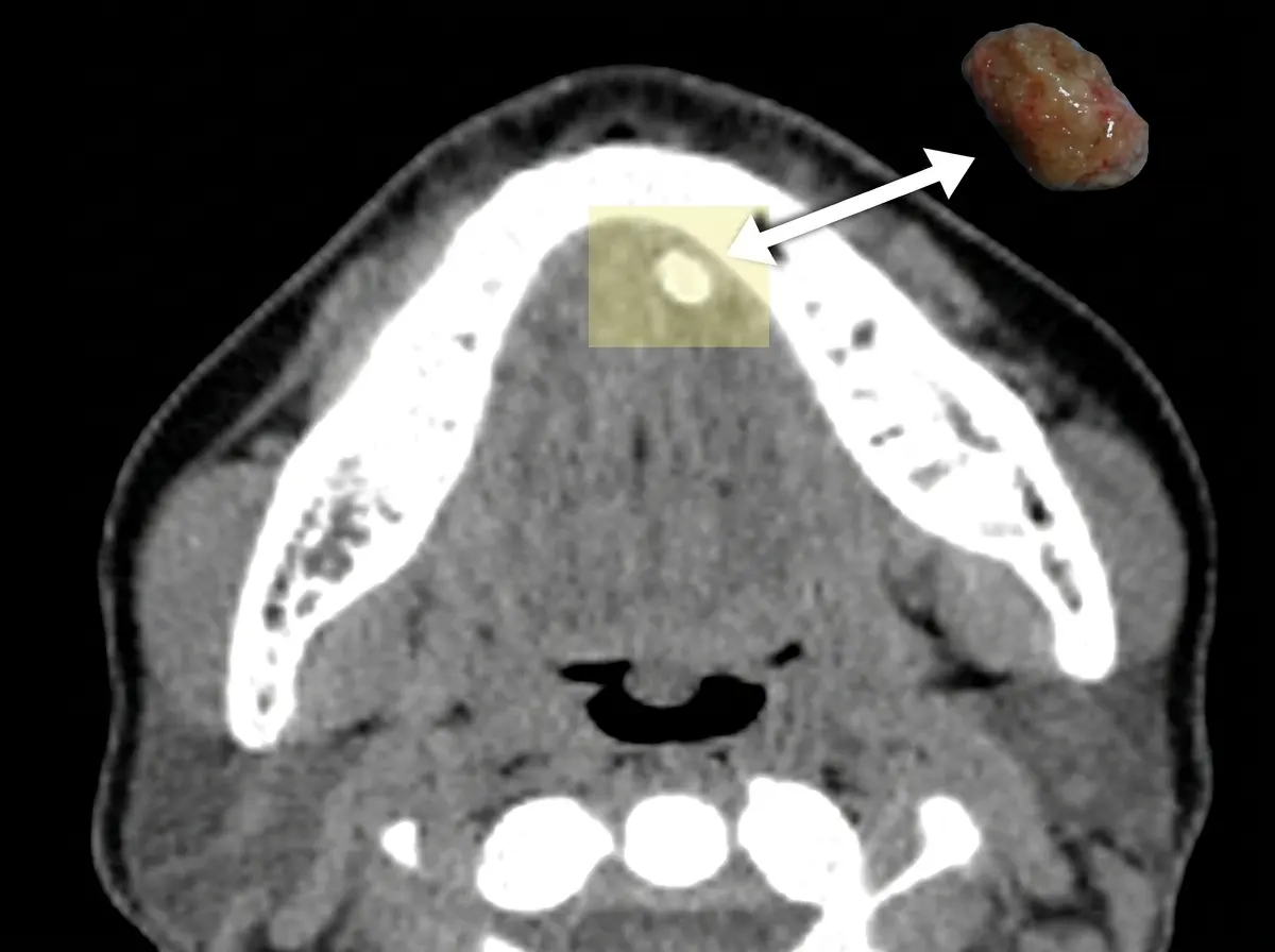
- CT scanning for calcified stones
- MRI in selected cases for soft tissue evaluation
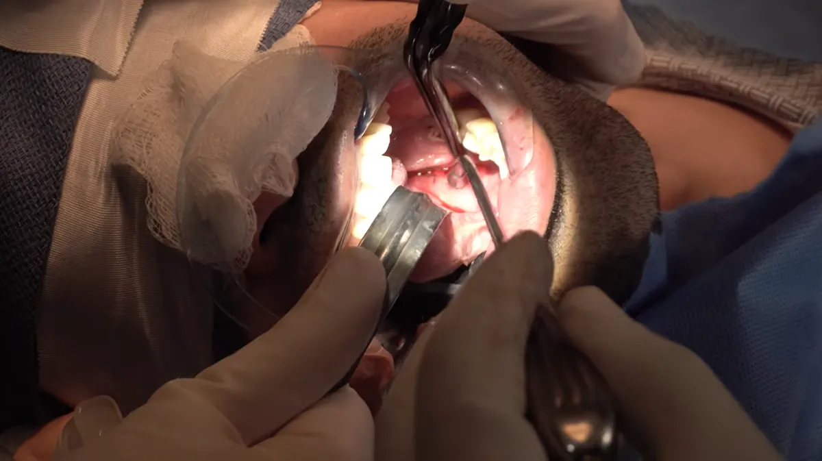
Diagnostic Sialendoscopy
Direct visualization of the ductal system allows both diagnosis and treatment in many cases.
Minimally Invasive First
Treatment of Salivary Gland Stones
Surgery remains the primary treatment, with sialendoscopy as the preferred first-line approach for most patients.
When open gland surgery is needed, Parotidectomy Recovery explains what to expect afterward, and Parotid Surgery Risks covers possible complications.
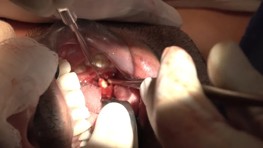
Sialendoscopy
Minimally invasive technique for direct visualization and removal of stones within the salivary ducts.
- Preservation of the salivary gland
- Minimal or no external incision
- Reduced recovery time
Combined Approaches
Larger or impacted stones may require combined endoscopic and limited open techniques.
Traditional Surgery
Complete gland removal is now reserved for rare cases where minimally invasive techniques are not feasible.
Our Approach
Treatment Philosophy
Gland Preservation First
The modern standard of care prioritizes maintaining salivary gland function whenever possible. Advances in endoscopic technology have dramatically reduced the need for gland excision.
Minimally Invasive Intervention
Early intervention allows for simpler procedures and improved outcomes, reducing the risk of chronic gland damage.
Case Gallery
Salivary Stone Library
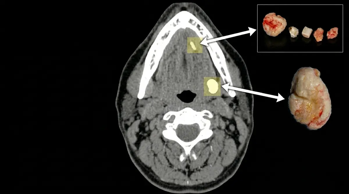
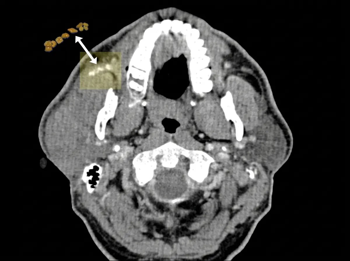
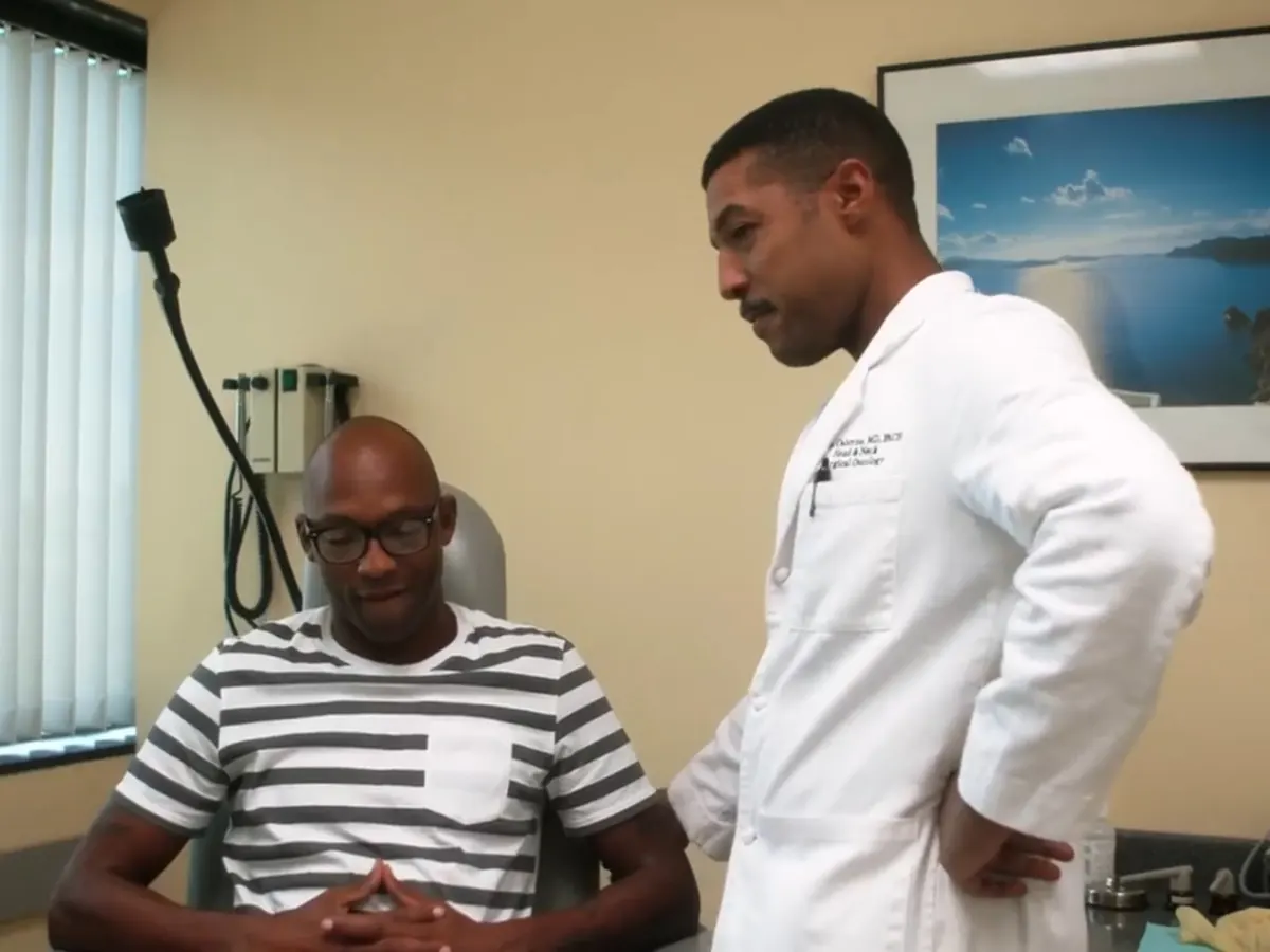
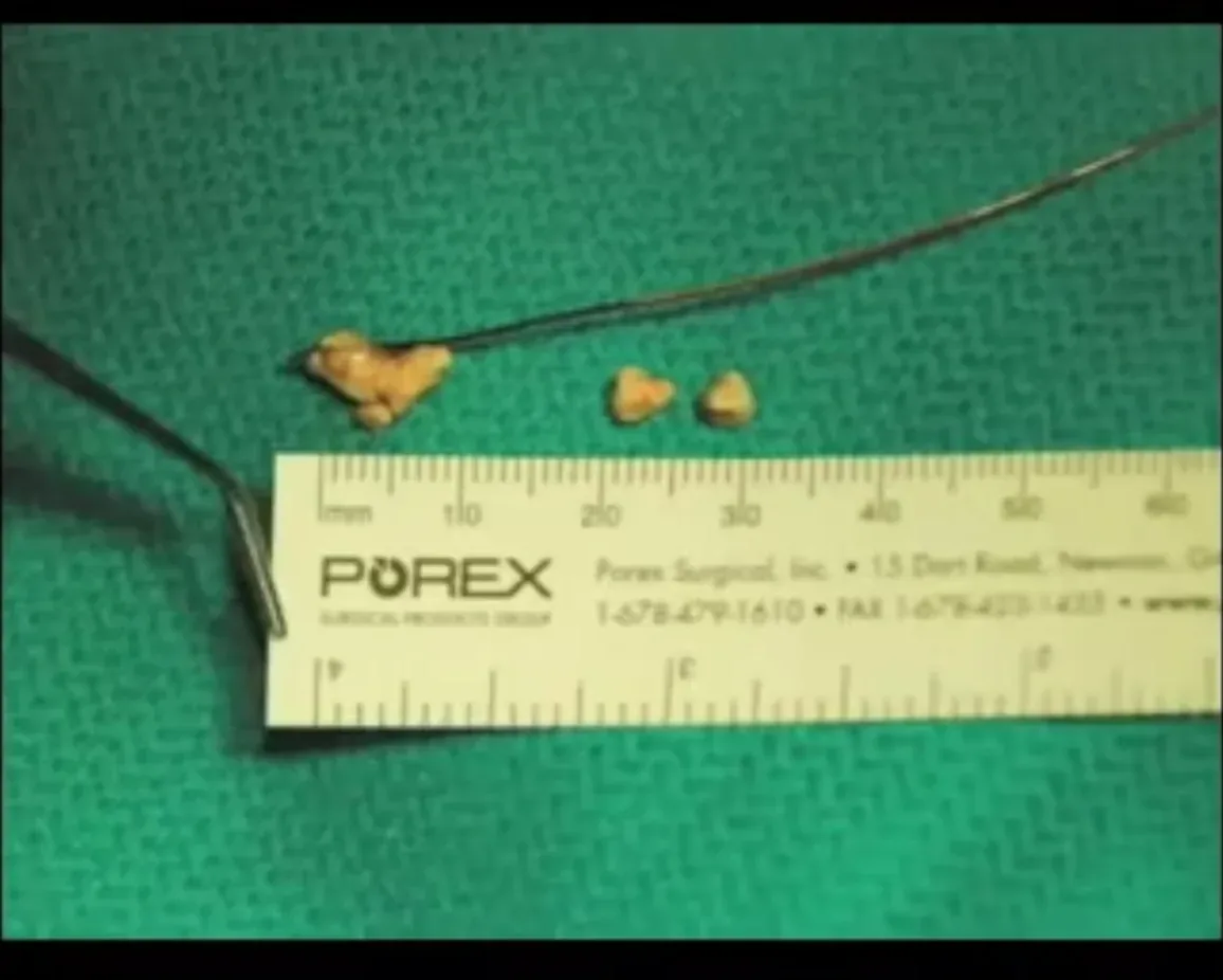
Documented sialendoscopy cases showing stones of varying size and number, each paired with the CT scan that located it before removal. Most of these patients were told their gland would have to be removed through the neck. All had their stones removed without external surgery.
Don't Wait
When to Seek Specialist Care
Evaluation by a specialist in salivary gland disorders is recommended for patients with:
Recurrent swelling associated with meals
Persistent gland discomfort
Prior infections or antibiotic use without resolution
Early diagnosis allows for less invasive treatment and improved long-term outcomes.
OHNI Case Files | Season 2: Episode 8
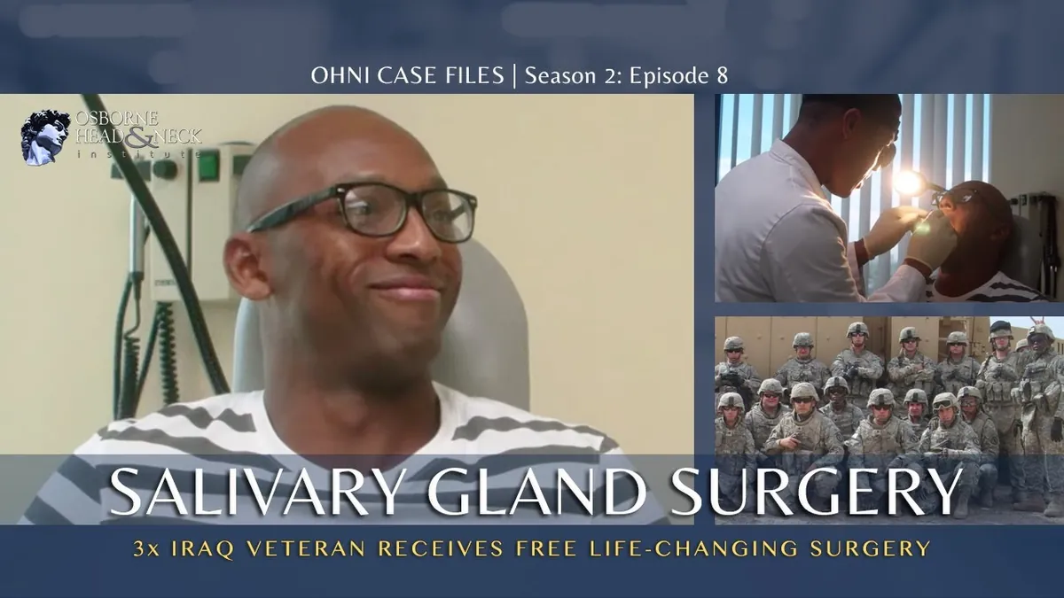
Watch: A Patient's Salivary Gland Surgery Story
A three-time Iraq veteran travelled to the Osborne Head & Neck Institute for treatment of a salivary gland condition. Follow his evaluation, surgery with Dr. Ryan F. Osborne, and recovery in this episode of OHNI Case Files.
Every case is planned around the individual patient, with surgery performed exclusively by Dr. Osborne.
Patients Treated for Salivary Gland Stones
"An ENT specialist in Perth told me I had a large stone in my submandibular gland and that the entire gland would need to be removed. Four opinions across Australia said the same. I am so happy I didn't take no for an answer and found an amazing surgeon who not only removed a very difficult and large stone, but also saved my gland."
M.G.
Australia
"My stone was very large and other doctors informed me my entire gland would need to be removed, resulting in a permanent scar and potential facial paralysis. Dr. Osborne's surgery was a smooth procedure without pain. I would definitely recommend Dr. Osborne and his wonderful staff."
L.B.
Maryland, United States
"I had a stone in my right salivary gland and was evaluating non-invasive treatment options. Dr. Osborne listened to my condition carefully and offered a treatment plan with no pressure to commit to surgery. Surgery was painless and the staff was super caring."
R.M.
California, United States
PATIENT EDUCATION
Salivary Gland Stone FAQs
Stones form when calcium and other minerals accumulate in the salivary ducts. Risk factors include dehydration, medications that reduce saliva flow, and chronic gland inflammation.
Small stones may sometimes pass spontaneously, but larger or impacted stones typically require treatment. Waiting can lead to infection and gland damage.
Sialendoscopy is a minimally invasive procedure that uses a tiny camera inserted into the salivary duct to locate and remove stones without external incisions.
Common signs include swelling and pain near the jaw during meals, which improves between meals. The pain is caused by saliva backing up behind the stone.
Recurrence is possible but uncommon with proper treatment. Staying hydrated and maintaining good oral health can help prevent new stones from forming.
Why Choose OHNI
Not Big Medicine | Personalized, Global Expertise
Many patients assume the best care is found at large, well-known institutions. While these centers offer excellent care, they often operate within standardized systems shaped by regional training models.
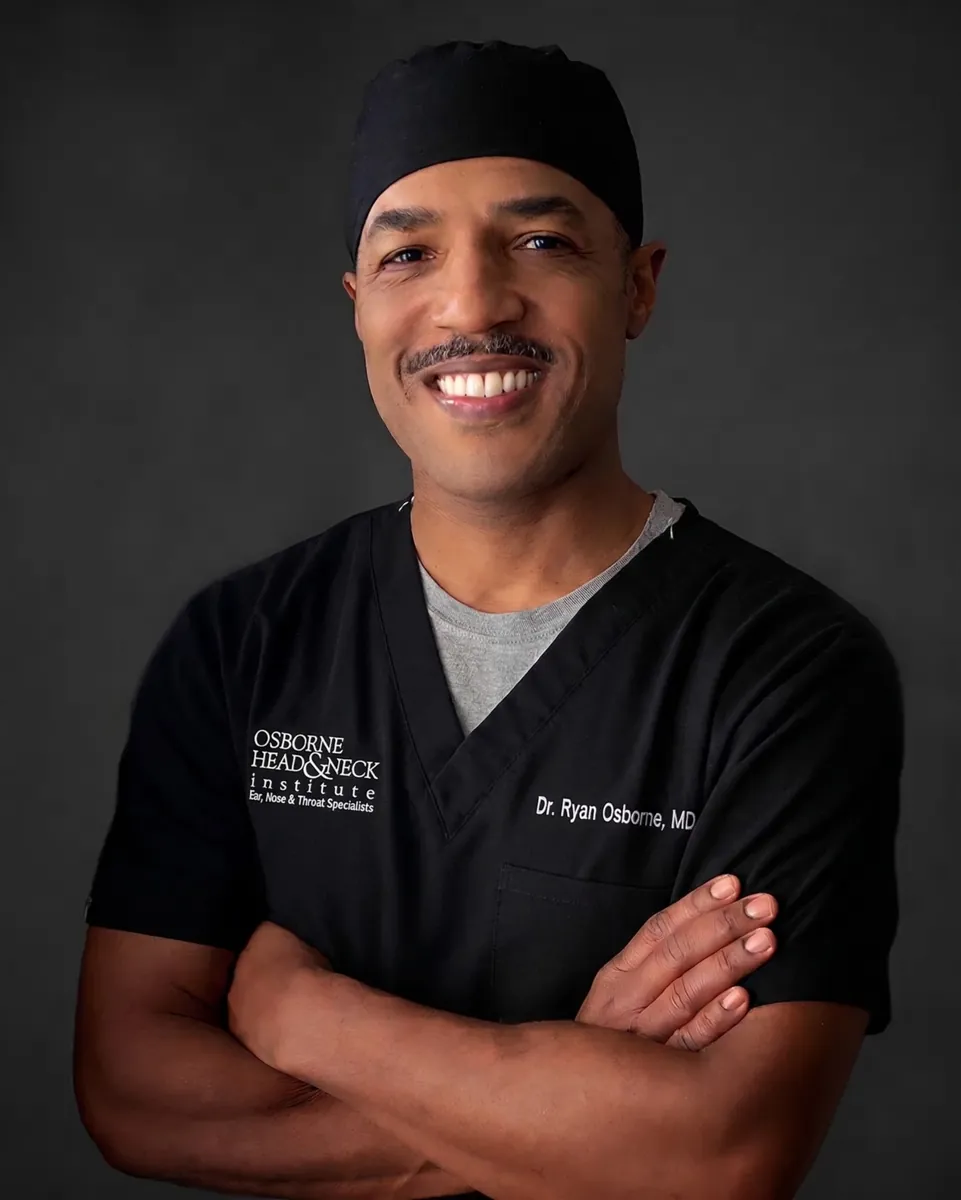
At OHNI, Dr. Ryan Osborne offers something fundamentally different: a truly global perspective combined with highly personalized surgical care with experience across five continents, tailoring each procedure to the individual patient, not the system.
Global Perspective
Experience across five continents, applying diverse surgical philosophies and techniques beyond standard U.S. training.
Precision & Preservation
Specialized techniques like Extra-Capsular Dissection designed to minimize invasiveness while protecting the facial nerve.
Patient-First Care
Direct surgeon access, individualized treatment planning, and a focus on both medical outcomes and quality of life.
Proven Outcomes
Patients consistently highlight clear communication, confident surgical decisions, and excellent functional results.
Expert Care for Salivary Gland Stones
With modern diagnostic techniques and minimally invasive therapies such as sialendoscopy, most patients can achieve complete symptom resolution while preserving normal gland function.
Become a Patient
Contact the Osborne Head & Neck Institute for expert evaluation of salivary gland stones and obstructive conditions.