Osborne Head & Neck Institute | Los Angeles
Salivary Stone Library
Sialendoscopy Cases | Imaging and Extracted Specimens
Ryan F. Osborne, MD | Fellowship-Trained Head & Neck Surgical Oncologist
Medically Reviewed By: OHNI Clinical Staff · Last Updated: November 4, 2026
About This Library
Stones Removed Without External Surgery
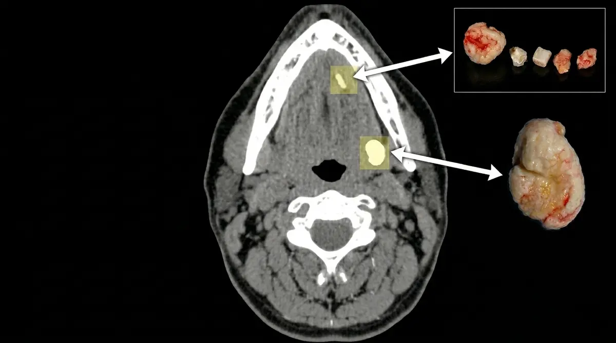
These are cases of sialendoscopy procedures performed on patients with salivary gland stones of varying sizes and quantities at the Osborne Head and Neck Institute. The CT scan (right) highlights the salivary gland, duct and stones. The stones are displayed (left) after removal from the patient, with a ruler to demonstrate size.
Most patients were told that their stones could not be removed without having neck surgery and removing the entire gland. All patients below had their stones removed without external surgery. Every procedure was performed by Dr. Ryan F. Osborne.
Case Gallery
Salivary Stone Extractions
Case 01
Extraction of Small Salivary Stones
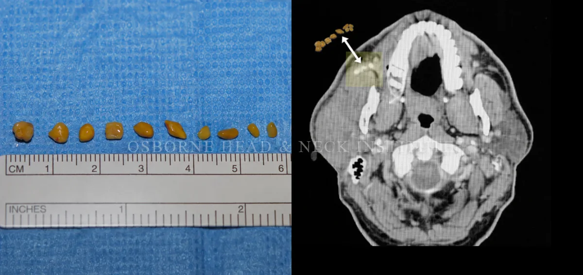
Figure: Stone, Axial CT, right parotid gland duct.
Case 02
Extraction of Small Salivary Stone
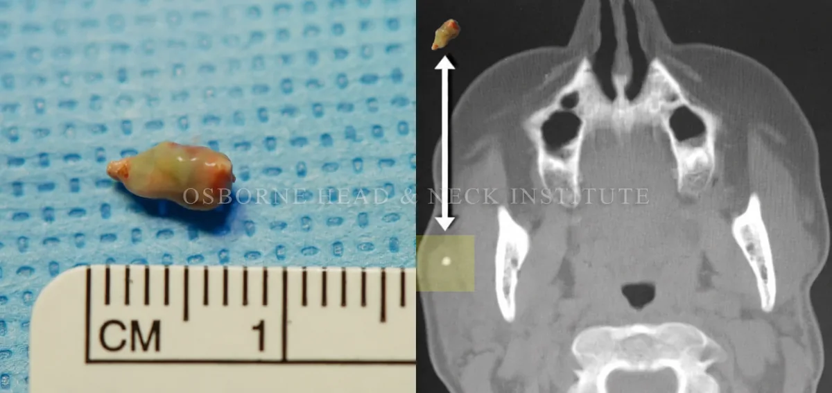
Figure: Stone, Axial CT, right parotid gland.
Case 03
Extraction of Small Salivary Stone
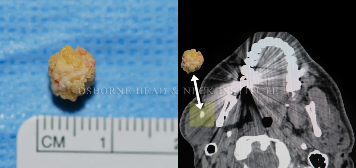
Figure: Stone, Axial CT, right parotid gland.
Case 04
Extraction of Small Salivary Stones
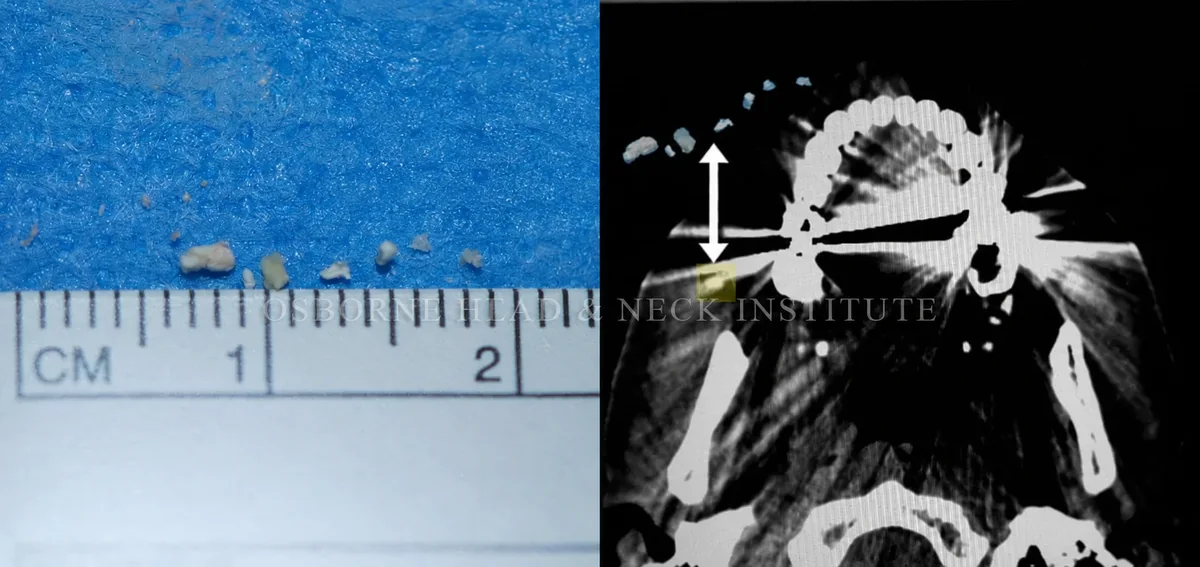
Figure: Stone, Axial CT, right parotid gland duct.
Case 05
Extraction of Small Salivary Stones
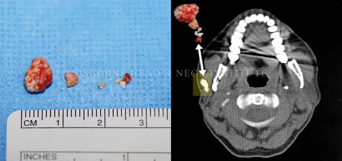
Figure: Stone, Axial CT, right parotid gland.
Case 06
Extraction of Large Salivary Stone
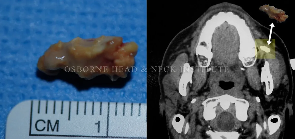
Figure: Stone, Axial CT, left parotid gland duct.
Case 07
Extraction of Small Salivary Stone
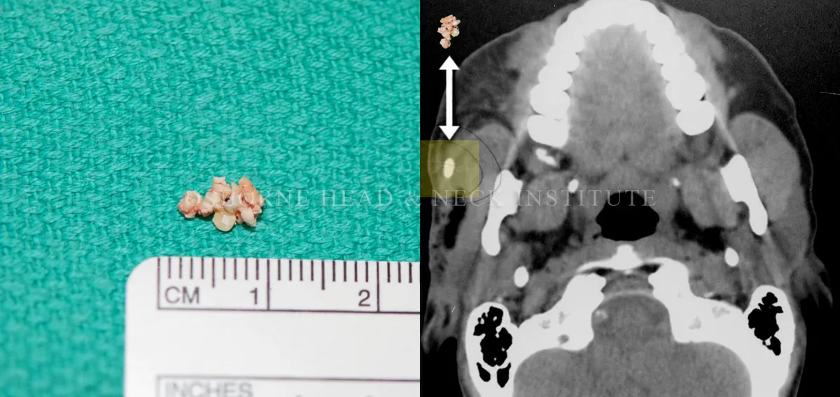
Figure: Stone, Axial CT, right parotid gland.
Case 08
Extraction of Small Salivary Stone
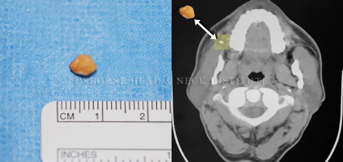
Figure: Stone, Axial CT, right parotid gland duct.
Case 09
Extraction of Small Salivary Stones
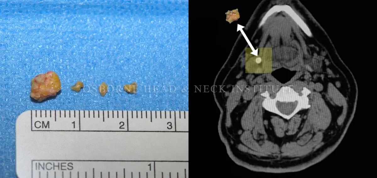
Figure: Stone, Axial CT, right submandibular gland.
Case 10
Extraction of Small Salivary Stone
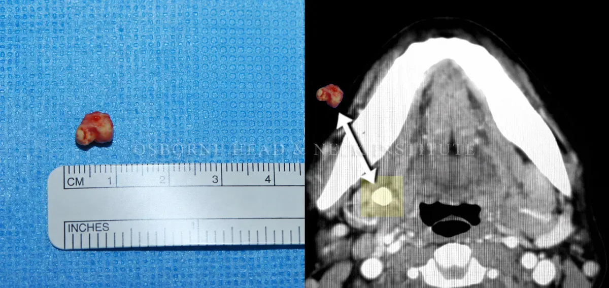
Figure: Stone, Axial CT, right submandibular gland.
Case 11
Extraction of Small Salivary Stones
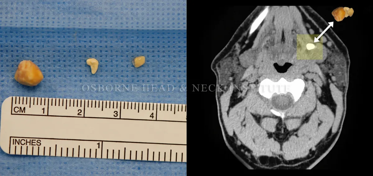
Figure: Stone, Axial CT, left submandibular gland.
Case 12
Extraction of Large Salivary Stone
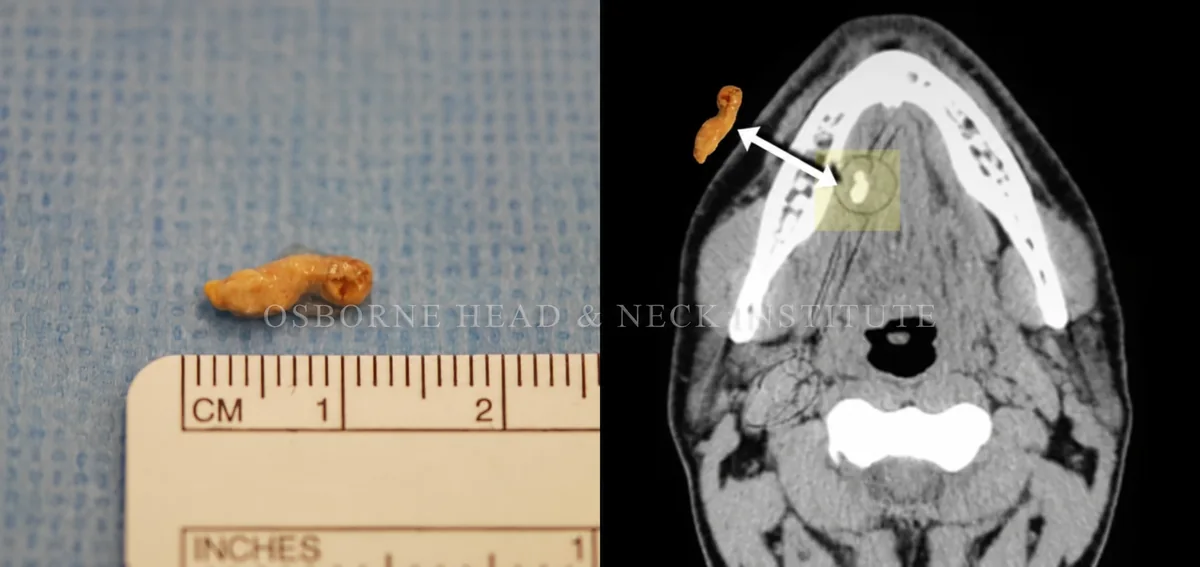
Figure: Stone, Axial CT, right submandibular gland duct.
Case 13
Extraction of Large and Small Salivary Stones
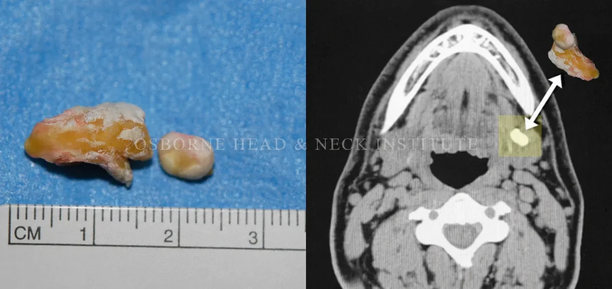
Figure: Stone, Axial CT, left submandibular gland.
Case 14
Extraction of Small Salivary Stones
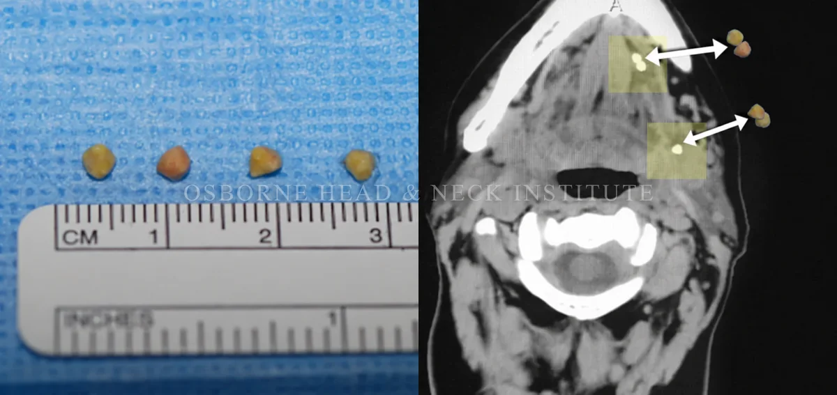
Figure: Stone, Axial CT, left submandibular gland and duct.
Case 15
Extraction of Small Salivary Stone
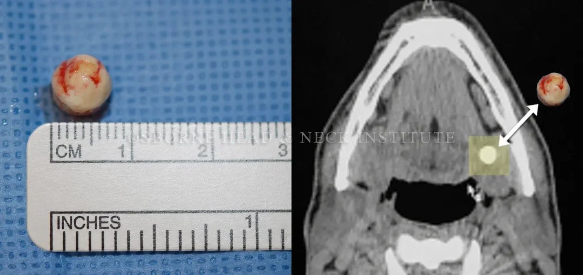
Figure: Stone, Axial CT, left submandibular gland.
Case 16
Extraction of Small Salivary Stone
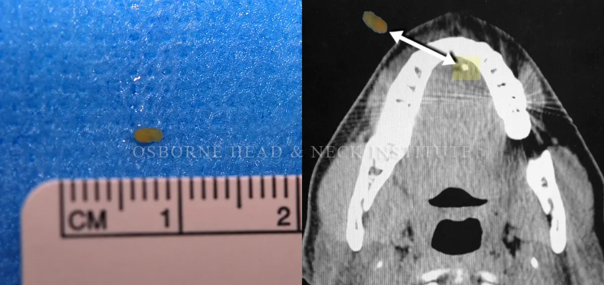
Figure: Stone, Axial CT, left submandibular gland duct.
Case 17
Extraction of Large Salivary Stone
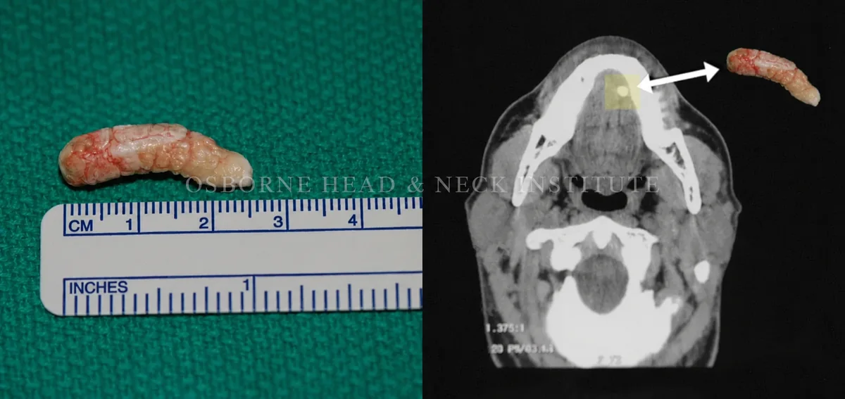
Figure: Stone, Axial CT, left submandibular gland duct.
Case 18
Extraction of Small Salivary Stones
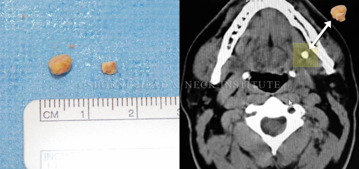
Figure: Stone, Axial CT, left submandibular gland.
Case 19
Extraction of Large and Small Salivary Stones
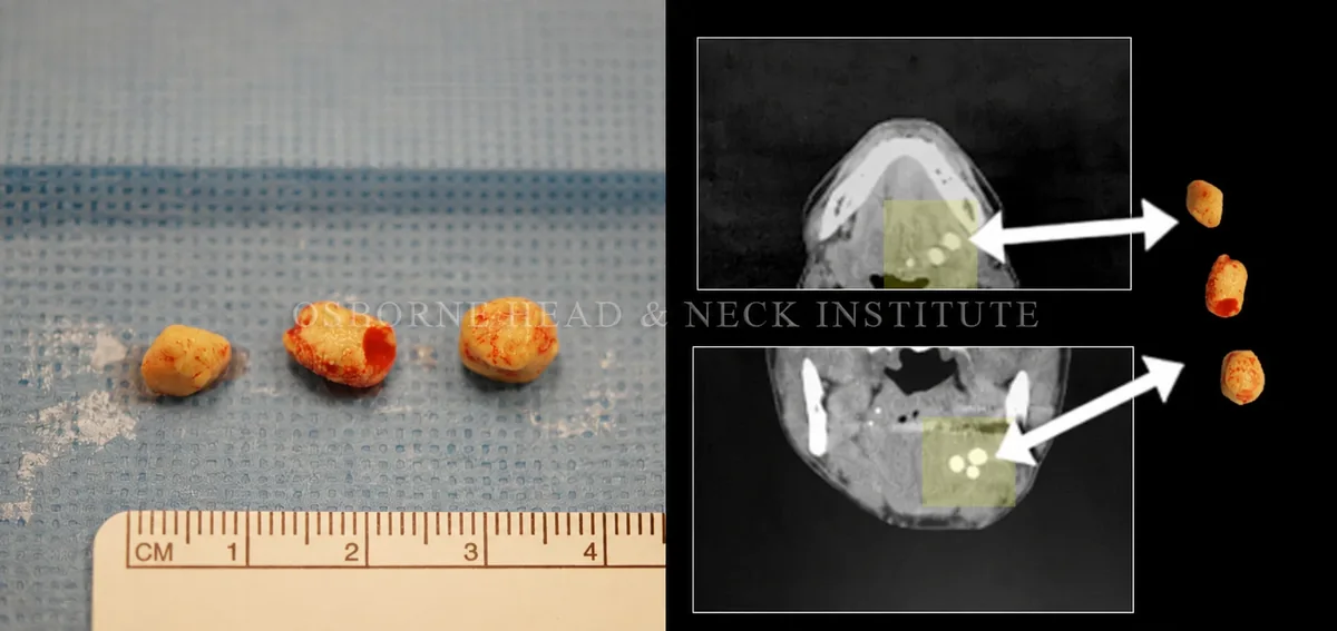
Figure: Stone, Axial CT (top) and Coronal CT (bottom), left submandibular gland.
Case 20
Extraction of Small Salivary Stone
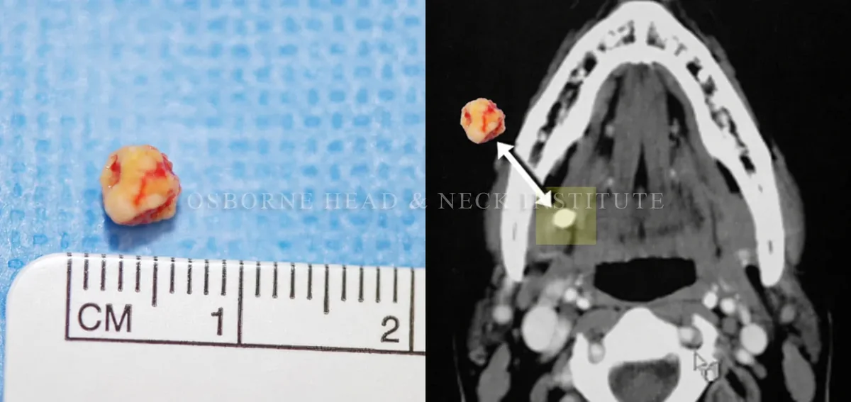
Figure: Stone, Axial CT, right submandibular gland.
Case 21
Extraction of Small Salivary Stone
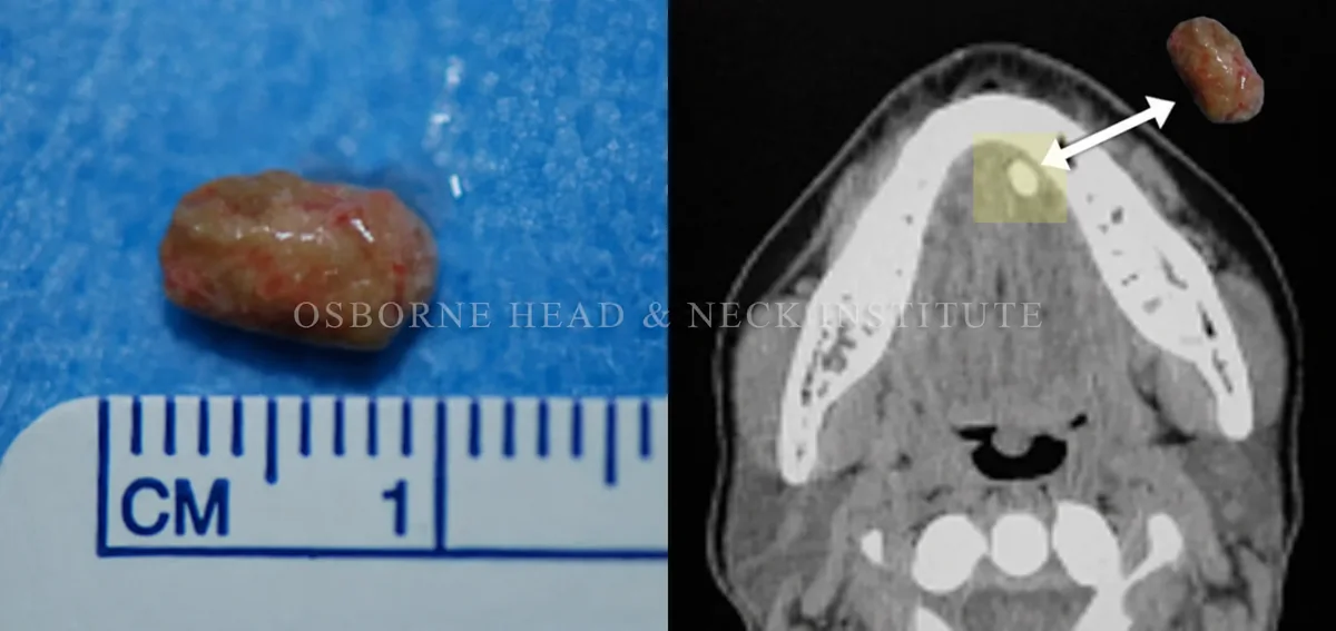
Figure: Stone, Axial CT, left submandibular gland duct.
Case 22
Extraction of Large and Small Salivary Stones
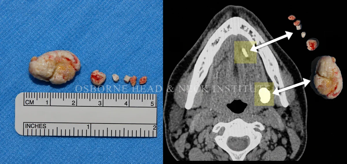
Figure: Stone, Axial CT, left submandibular gland and duct.
Expert Care for Salivary Gland Stones
If you have been told your salivary gland must be removed because of a stone, a second opinion with a salivary gland specialist may reveal a minimally invasive, gland-preserving option.
Become a Patient
Contact the Osborne Head & Neck Institute for expert evaluation of salivary gland stones and obstructive conditions.