Swelling of a salivary gland during meals is a distinctive and often concerning symptom. Many patients describe a pattern in which the gland becomes enlarged or uncomfortable while eating, only to partially improve afterward. This cyclical swelling is not random, it is a hallmark sign of obstructive salivary gland disease, most commonly caused by salivary gland stones (sialolithiasis).1,3
Although this condition is typically benign, it can significantly impact quality of life and may lead to recurrent infections or chronic gland damage if left untreated. Understanding why this symptom occurs, what it means, and how it is treated is essential for timely and effective care.4
Understanding Salivary Flow: Why Symptoms Occur During Eating
Salivary glands produce saliva continuously, but production increases significantly during meals. This is a normal physiologic response that supports digestion and lubrication.
The three major salivary glands include:
- Parotid glands (near the ear)
- Submandibular glands (beneath the jaw)
- Sublingual glands (under the tongue)
When functioning normally, saliva flows through ducts into the mouth without resistance. However, when a blockage is present, saliva cannot exit freely. This results in increased pressure within the gland, swelling and discomfort, and temporary enlargement during meals.
The Most Common Cause: Salivary Gland Stones (Sialolithiasis)
What Are Salivary Stones?
Salivary gland stones are calcified deposits that form within the salivary ducts. They are the most common cause of obstructive salivary gland disease. These stones can partially block saliva flow, completely obstruct the duct, or cause intermittent or persistent symptoms.1,4
Why Do Stones Cause Swelling With Eating?
During meals, saliva production increases. If a stone blocks the duct, saliva accumulates behind the obstruction, pressure builds within the gland, and the gland becomes swollen and painful. Once the stimulus (eating) ends, swelling may partially subside as pressure decreases.
This pattern people experience, their jaw and face swelling worsens with eating, is highly characteristic of salivary gland stones.
Which Gland Is Most Commonly Affected?
The submandibular gland is most frequently affected due to thicker saliva, upward ductal flow, and a more alkaline environment. However, the parotid gland can also develop stones, often with more subtle or complex symptoms.
Other Causes of Swelling With Eating
1. Ductal Narrowing (Stricture)
Narrowing of the salivary duct can restrict flow even without a stone. This may result from prior inflammation and can lead to recurrent swelling.
2. Chronic Parotitis
Chronic inflammation of the gland may impair saliva flow, causing recurrent swelling often associated with ductal abnormalities.
3. Autoimmune Conditions
Conditions such as Sjögren's syndrome can affect gland function, causing reduced saliva production, intermittent swelling, and associated dry mouth.
Symptoms Associated With Salivary Obstruction
Patients with obstructive salivary gland disease may experience:
- Swelling during meals
- Pain or pressure in the gland
- Recurrent episodes of enlargement
- Reduced saliva flow
- Occasional infection
In more advanced cases, persistent swelling and fever or redness (infection) may develop.
How Is Salivary Gland Obstruction Diagnosed?
Clinical Evaluation
Initial assessment includes symptom pattern (especially meal-related swelling), physical examination of the gland, and evaluation of duct openings.
Imaging
Imaging helps confirm the diagnosis and locate the obstruction:
- Ultrasound: first-line for many cases
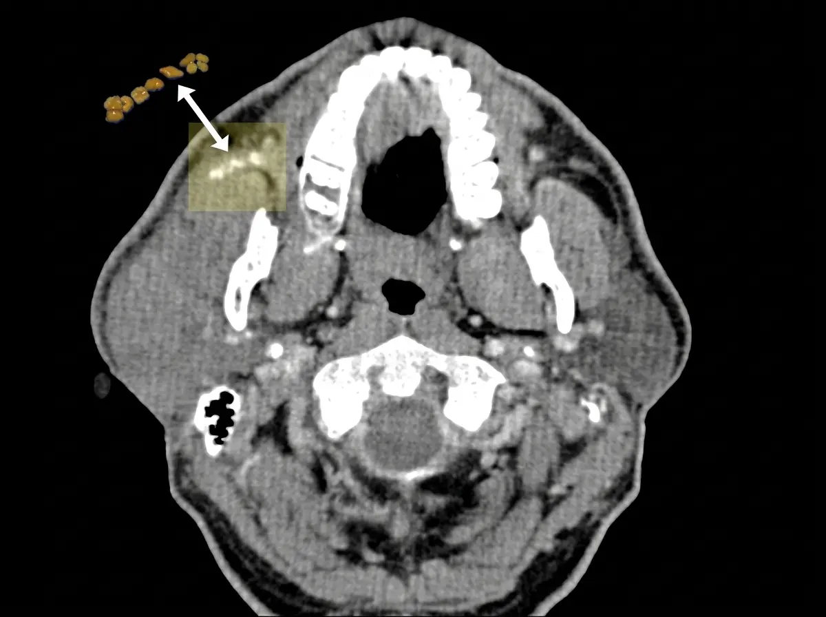
- CT scan: highly effective for detecting stones
- MRI: useful in complex cases
Diagnostic Sialendoscopy
In many cases, diagnosis and treatment can be combined using sialendoscopy, a minimally invasive technique that allows direct visualization of the duct system.
Treatment Options for Salivary Gland Stones
1. Sialendoscopy (Primary Treatment)
Sialendoscopy is now the preferred treatment for most salivary stones. It is minimally invasive with no external incision, preserves the gland, and allows rapid recovery. Small stones can often be removed directly through the duct.1,2
If open gland surgery is needed, Parotidectomy Recovery explains what to expect afterward.
2. Combined Techniques
For larger stones, endoscopic combined with small incision approaches are used, still designed to preserve the gland.
3. Traditional Surgery (Rare)
Removal of the entire gland is now uncommon and reserved for complex cases.
How This Differs From a Tumor
Patients often confuse swelling with eating and a lump near the jaw. Key differences:
| Feature | Salivary Stone | Parotid Tumor |
|---|---|---|
| Timing | During meals | Persistent |
| Pain | Common | Often painless |
| Pattern | Intermittent | Progressive |
Why Early Treatment Matters
Delaying treatment may lead to recurrent infections, chronic gland damage, progressive ductal scarring, and more invasive procedures. Early intervention allows for simpler procedures, better outcomes, and preservation of gland function.2,3
When to Seek Medical Attention
You should seek evaluation if you experience:
- Recurrent swelling during meals
- Persistent gland enlargement
- Pain or infection
- Difficulty eating due to swelling
Early evaluation allows for minimally invasive treatment and prevents complications.
Summary
Swelling of a salivary gland during eating is a classic sign of obstructive salivary gland disease, most often caused by salivary stones. While typically benign, this condition can lead to recurrent symptoms and complications if left untreated.
Accurate diagnosis and modern treatment, particularly sialendoscopy, allow for effective management while preserving gland function. With early intervention, most patients achieve complete symptom resolution and return to normal function.