Salivary gland stones (sialolithiasis) are the most common cause of obstructive salivary gland disease and frequently present with swelling, pain, and discomfort, especially during meals. While treatment has traditionally involved surgical removal of the gland, advances in minimally invasive techniques have transformed management.1,3
Today, sialendoscopy has emerged as the preferred first-line treatment in many cases, offering gland preservation and faster recovery. However, traditional surgery still plays an important role in selected situations. Understanding the differences between these approaches is essential for determining the most appropriate treatment strategy.1,2
Understanding Salivary Gland Stones
Salivary stones form within the ducts of the salivary glands and block the normal flow of saliva. This leads to swelling during eating, pain or pressure in the gland, and recurrent infections.3
Sialolithiasis most commonly affects the submandibular gland, but the parotid gland may also be involved.3
Traditional Surgery: The Historical Approach
Before the development of minimally invasive techniques, treatment often involved removal of the affected salivary gland.
Types of Surgery
- Submandibular gland excision for stones in the submandibular gland
- Parotidectomy for parotid gland disease
Advantages
- Definitive removal of the diseased gland
- Effective for large or inaccessible stones
- Long-standing, well-established approach
Disadvantages
- External incision and visible scar
- Risk to surrounding structures
- Risk of facial paralysis
- Longer recovery time
- Permanent loss of gland function
Sialendoscopy: A Minimally Invasive Alternative
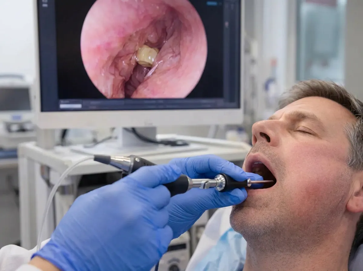
Sialendoscopy is an advanced technique that uses a small endoscope to access the salivary duct internally. This allows for direct visualization of the duct system, identification of stones or narrowing, and simultaneous treatment.1
Benefits of Sialendoscopy
- No external incision
- Preservation of the salivary gland
- Shorter recovery time
- No risk of facial paralysis
Studies demonstrate high success rates and significant symptom improvement with this approach.2
When Is Sialendoscopy the Best Option?
Sialendoscopy is most effective for:
- Small to medium-sized stones
- Stones located within accessible ducts
- Ductal narrowing (strictures)
- Recurrent swelling without large obstruction
When Is Surgery Still Necessary?
Large or Impacted Stones
Stones that are too large to remove endoscopically or deeply embedded within the gland may require a minimally invasive combined approach.
Recurrent or Severe Disease
Patients with chronic gland damage, repeated infections, or failed prior treatments may benefit from gland removal.
Complex Anatomy
In some cases, ductal anatomy may limit access for endoscopic techniques.
Combined Approaches: The Modern Standard
In many cases, treatment is not strictly one or the other. Hybrid techniques include:
- Endoscopic guidance with small incision
- Fragmentation of large stones
- Targeted removal with gland preservation
These approaches combine the strengths of both methods while minimizing risks.
Comparing Sialendoscopy and Surgery
| Feature | Sialendoscopy | Traditional Surgery |
|---|---|---|
| Incision | None | External incision |
| Gland preservation | Yes | No |
| Recovery time | Short | Longer |
| Risk to nerves | 0% | 0–11% |
| Ideal for | Small–medium stones | Large/complex stones |
Recovery Differences
Sialendoscopy Recovery
- Return to normal activity within 24 hours
- Minimal discomfort
- No visible scarring
Surgical Recovery
- 7 - 10 days for recovery
- Temporary swelling and discomfort
- Potential cosmetic considerations
Risks and Complications
Sialendoscopy Risks
- Mild swelling
- Temporary discomfort
- Rare ductal injury
Surgical Risks
- Facial nerve weakness (parotid surgery)
- Scarring
- Infection or bleeding
Why Treatment Selection Matters
Choosing the appropriate treatment approach impacts long-term gland function, recovery experience, and risk of recurrence. Modern care emphasizes minimally invasive treatment first and gland preservation whenever possible.1,2
The Role of Specialist Care
Effective management of salivary gland stones requires accurate diagnosis, advanced imaging, and expertise in both endoscopic and surgical techniques. Specialists trained in salivary gland disorders can determine whether sialendoscopy is appropriate and when combined or surgical approaches are needed.
When to Seek Treatment
You should seek evaluation if you experience:
- Swelling during meals
- Recurrent gland pain
- Persistent symptoms despite conservative care
Early evaluation allows for less invasive treatment options.
Summary
Sialendoscopy and traditional surgery represent two important approaches to treating salivary gland stones. While surgery was historically the standard, sialendoscopy has transformed care by offering a minimally invasive, gland-preserving alternative.
Most patients can now be treated effectively without gland removal, though surgery remains important for complex cases. A tailored approach based on stone size, location, and patient factors ensures the best outcomes.