
Osborne Head & Neck Institute | Los Angeles
Salivary Gland Cysts (Sialocele)
Understanding Salivary Fluid Accumulation After Trauma or Surgery
Ryan F. Osborne, MD, FACS | Fellowship-Trained Head & Neck Surgical Oncologist
Medically Reviewed By: OHNI Clinical Staff · Last Updated: November 3, 2026
Overview
What Is a Salivary Gland Cyst (Sialocele)?
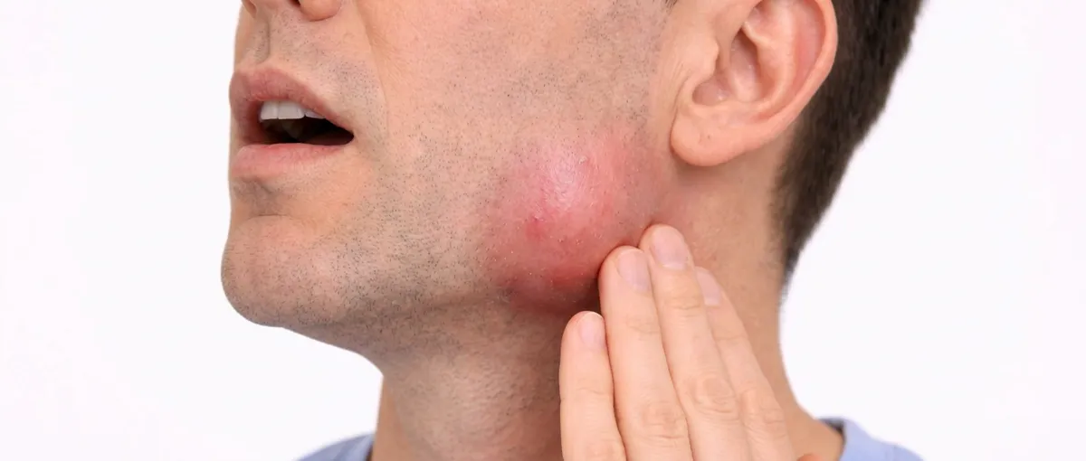
A sialocele is a fluid-filled collection of saliva that forms when normal salivary flow is disrupted. Unlike true cysts lined by epithelium, it represents an accumulation of saliva within tissues from injury or disconnection between the gland and its ductal system.
How It Forms
Saliva travels through a network of ducts into the oral cavity. When this pathway is interrupted, saliva can leak into surrounding tissues or remain trapped within the gland, forming a cyst-like structure.
Most Commonly Associated With
Surgical procedures involving the salivary glands
Physical trauma to the face or neck
Less commonly, spontaneous obstruction of salivary ducts
Anatomy
Understanding the Salivary System
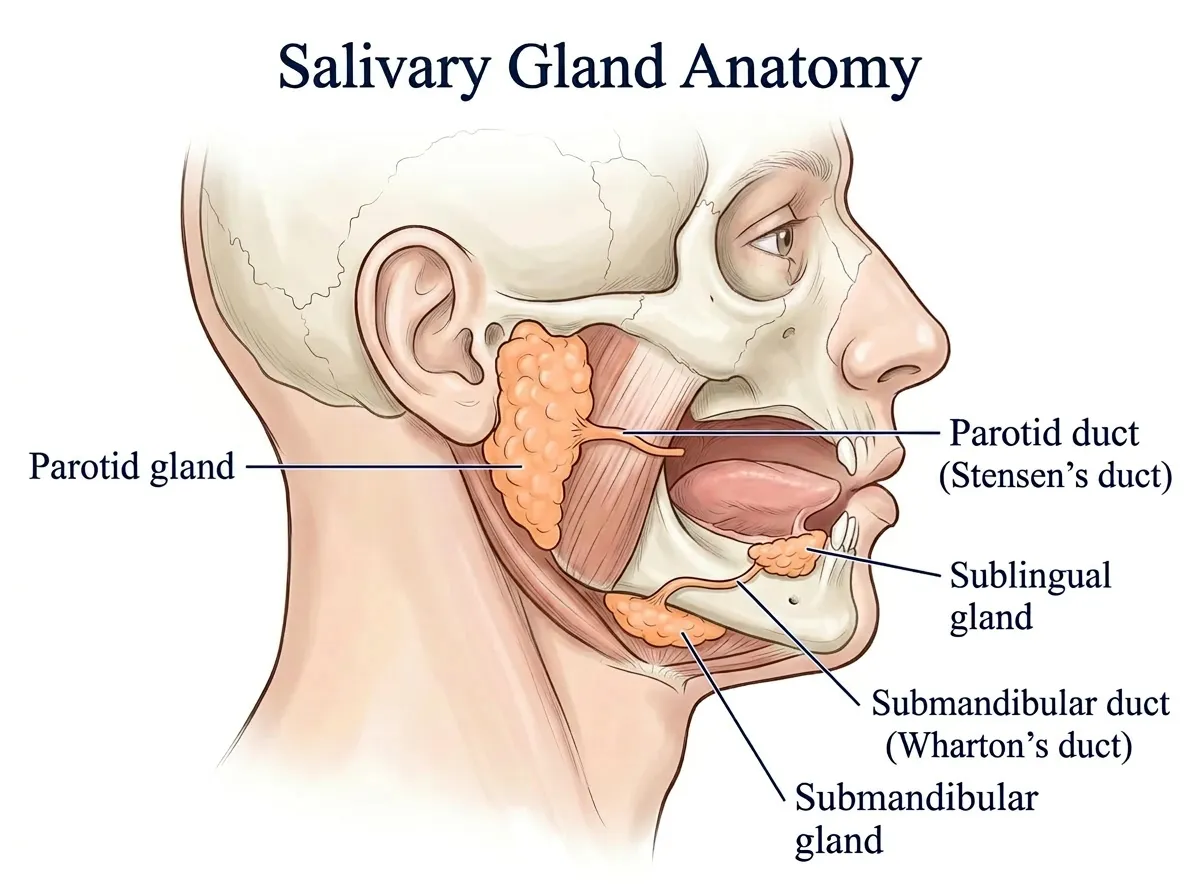
The salivary glands play a critical role in oral health and digestion. They produce saliva, which lubricates the mouth, initiates digestion, protects teeth and mucosa, and helps with speech and swallowing.
The parotid gland, the largest salivary gland, is particularly prone to sialocele formation due to its size and anatomical complexity. Each gland is connected to the mouth through ducts. When these ducts are damaged or obstructed, saliva cannot exit normally, leading to accumulation and cyst formation.
Pathogenesis
How a Sialocele Forms
The development of a sialocele follows a predictable process resulting in a persistent fluid collection.
Duct Disruption
Trauma or surgery interrupts the connection between gland and duct
Saliva Leakage
Saliva escapes into surrounding tissues or becomes trapped
Fluid Buildup
Continuous saliva production leads to progressive accumulation
Cyst Formation
A cyst-like cavity forms as saliva accumulates faster than reabsorbed
Etiology
Causes of Sialocele
1. Surgical Trauma
The most frequent cause. During surgery, small ductal branches may be disrupted, leaking saliva postoperatively.
- Parotidectomy
- Removal of salivary gland tumors
- Neck or facial surgery
2. Physical Trauma
Injury to the face or jaw can damage salivary glands or ducts.
- Motor vehicle accidents
- Sports injuries
- Falls or blunt trauma
- Facial lacerations
3. Spontaneous Occurrence
In some cases, sialoceles develop without a clear cause. Possible contributing factors include:
- Duct obstruction
- Inflammation
- Increased salivary pressure
Clinical Presentation
Symptoms of Salivary Gland Cysts
Sialoceles are typically benign but can present with a range of symptoms depending on size and location.
Common Symptoms
- Soft swelling in the cheek, jaw, or neck
- Gradual enlargement of the affected area
- Fluctuating size (may increase with eating)
- Pain or pressure due to tissue stretching
- Feeling of fullness
Functional Effects
- Chewing difficulties
- Speaking impairment
- Swallowing interference
Potential Complications
- Infection due to stagnant saliva
- Skin breakdown or fistula formation
- Cosmetic deformity
Evaluation
Diagnosis of Salivary Gland Cysts
Clinical Evaluation
Diagnosis is typically based on:
- Patient history (recent surgery or trauma)
- Physical examination
- Location and characteristics of swelling
Imaging Studies
In certain cases, imaging may be used to confirm diagnosis:
- Ultrasound
- CT scan
- MRI
These studies help define the extent of the fluid collection, identify ductal injury, and rule out other conditions.
Expertise
Why Specialist Care Matters
Sialoceles are frequently mismanaged when treated without addressing the underlying cause. A salivary gland specialist focuses on three critical principles:
Precise Treatment
Ensuring that any underlying pathology is fully addressed
Anatomical Management
Careful handling of remaining salivary tissue and ducts
Prevention of Recurrence
Using advanced techniques to prevent continued saliva leakage
Prevention
Prevention of Sialocele
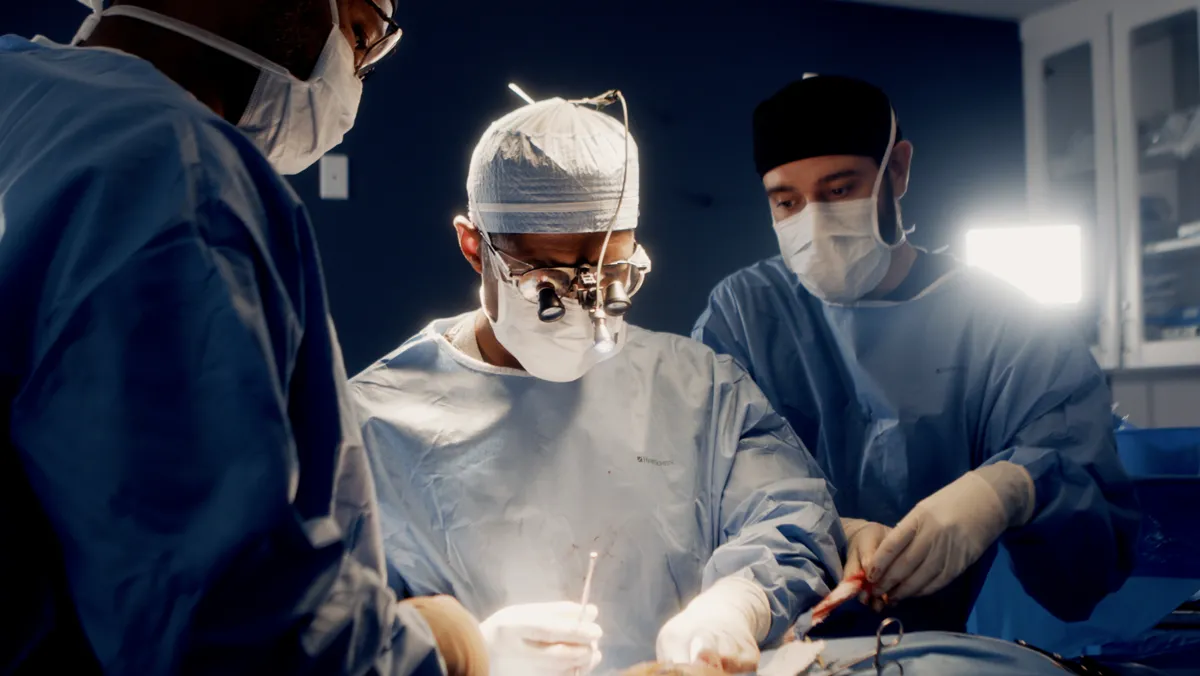
The most effective way to prevent sialocele formation is through meticulous surgical technique and proper postoperative care.
Surgical Technique
- Careful dissection around delicate structures
- Identification of ductal branches
- Sealing or ligating disrupted ducts
- Preservation of surrounding tissues
Reconstruction & Closure
- Identify and seal residual ductal pathways
- Prevent saliva leakage into surrounding tissue
- Reconstruct the surgical bed when necessary
Postoperative Care
- Placement of surgical drains
- Use of compressive dressings
- Close follow-up monitoring
- Dietary modifications to reduce saliva production
Treatment
Treatment of Sialocele
Simple drainage provides temporary relief but does not address the source of leakage. Effective treatment focuses on stopping the source.
Sialoceles can follow parotid surgery; Parotid Surgery Risks and Parotidectomy Recovery explain how healing usually progresses.
Repeated Aspiration
Used for initial symptom relief, often combined with other therapies.
Pressure Dressings
External compression helps reduce fluid accumulation during healing.
Saliva Suppression
Medications or dietary changes may reduce saliva production during healing.
Botulinum Toxin (Botox)
Temporarily reduces salivary gland activity, allowing the ductal system to heal.
Surgical Management
Repair of ductal injury, removal of affected gland tissue, or reconstruction for definitive resolution.
Recurrence
Risk of Recurrence
Sialoceles have a high recurrence rate when not properly treated. Specialist-directed care significantly reduces recurrence risk.
Factors That Increase Recurrence
- Incomplete treatment
- Failure to address ductal disruption
- Continued saliva production
- Inadequate postoperative care
Risk Factors
Who Is at Risk?
Higher risk individuals include:
- Patients undergoing salivary gland surgery
- Individuals with facial trauma
- Patients with prior salivary gland disease
- Those with complex or extensive surgical procedures
PATIENT EDUCATION
Salivary Gland Cyst FAQs
A sialocele is a fluid-filled collection of saliva caused by disruption of normal salivary flow.
Most commonly: surgical trauma, physical injury, or duct obstruction.
Some may resolve, but many persist or recur without proper treatment.
Yes. Drainage alone does not treat the underlying cause, making recurrence likely.
A salivary gland specialist is best equipped to provide definitive treatment.
Philosophy
Treatment Philosophy
Sialocele care is not about removing fluid, it is about identifying the source, restoring anatomy, and preventing recurrence.
Precision Over Temporary Solutions
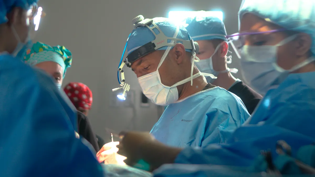
Identify the source, restore anatomy, prevent recurrence, never settle for drainage alone.
Experience Matters
Outcomes depend on surgical expertise, anatomical mastery, and meticulous attention to detail.
Prevention Is the Best Treatment
Advanced technique, careful reconstruction, and disciplined postoperative care prevent recurrence.
Why Choose OHNI
Not Big Medicine | Personalized, Global Expertise
Many patients assume the best care is found at large, well-known institutions. While these centers offer excellent care, they often operate within standardized systems shaped by regional training models.
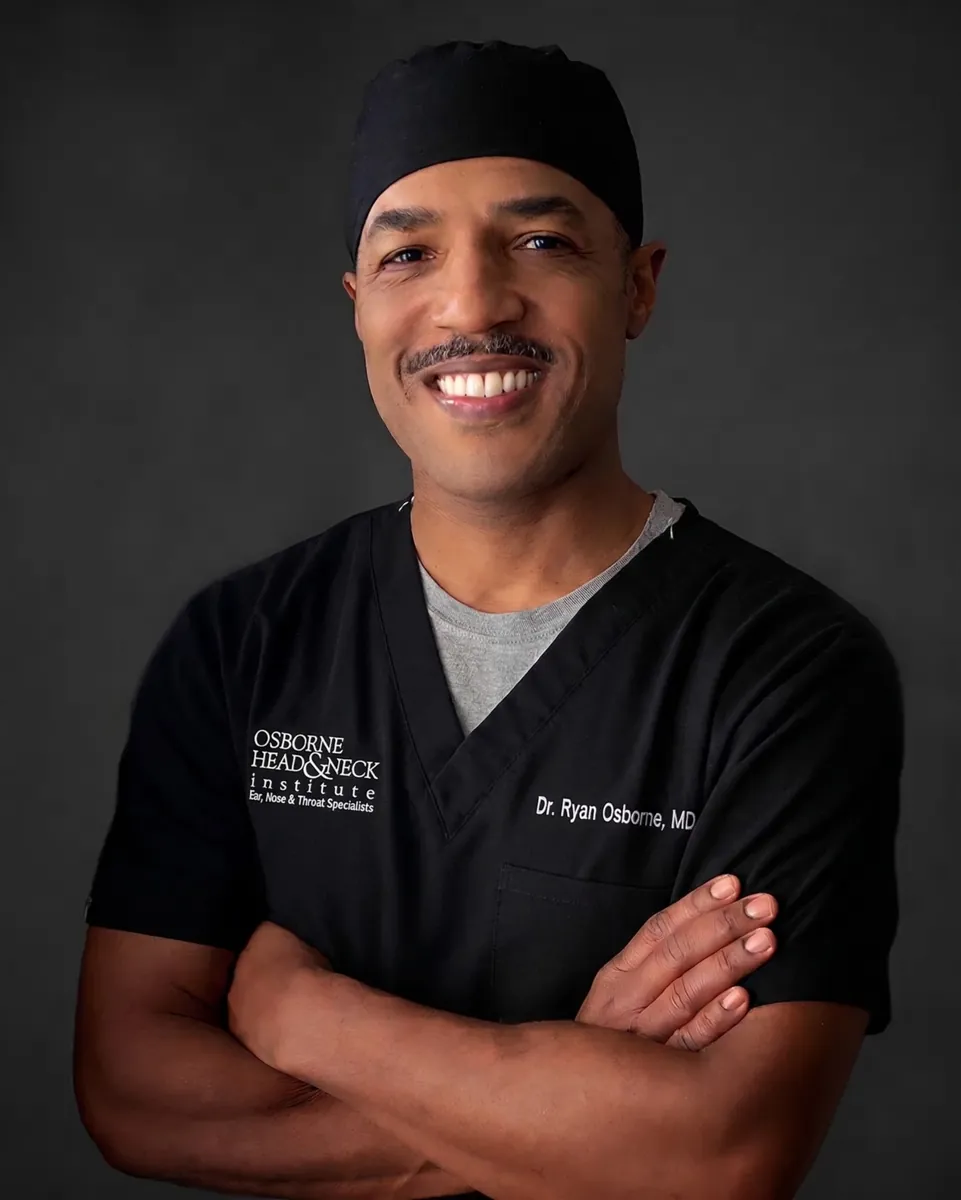
At OHNI, Dr. Ryan Osborne offers something fundamentally different: a truly global perspective combined with highly personalized surgical care with experience across five continents, tailoring each procedure to the individual patient, not the system.
Global Perspective
Experience across five continents, applying diverse surgical philosophies and techniques beyond standard U.S. training.
Precision & Preservation
Specialized techniques like Extra-Capsular Dissection designed to minimize invasiveness while protecting the facial nerve.
Patient-First Care
Direct surgeon access, individualized treatment planning, and a focus on both medical outcomes and quality of life.
Proven Outcomes
Patients consistently highlight clear communication, confident surgical decisions, and excellent functional results.
Take Action
When to Seek Evaluation
You should seek evaluation if you experience:
- Persistent swelling after salivary gland surgery
- Facial swelling following trauma
- Recurrent fluid collections
- Pain or signs of infection
Early intervention for salivary gland cysts leads to better outcomes and reduces complications.